1K1W

Crystal structure of 4-alpha-glucanotransferase from thermococcus litoralis


Experimental Data Snapshot

  • Method: X-RAY DIFFRACTION
  • Resolution: 2.80 Å
  • R-Value Free: 0.289 
  • R-Value Work: 0.221 
  • R-Value Observed: 0.224 

wwPDB Validation   3D Report Full Report


Ligand Structure Quality Assessment 


This is version 1.3 of the entry. See complete history


Literature

Crystal structures of 4-alpha-glucanotransferase from Thermococcus litoralis and its complex with an inhibitor

Imamura, H.Fushinobu, S.Yamamoto, M.Kumasaka, T.Jeon, B.S.Wakagi, T.Matsuzawa, H.

(2003) J Biol Chem 278: 19378-19386

  • DOI: https://doi.org/10.1074/jbc.M213134200
  • Primary Citation of Related Structures:  
    1K1W, 1K1X, 1K1Y

  • PubMed Abstract: 

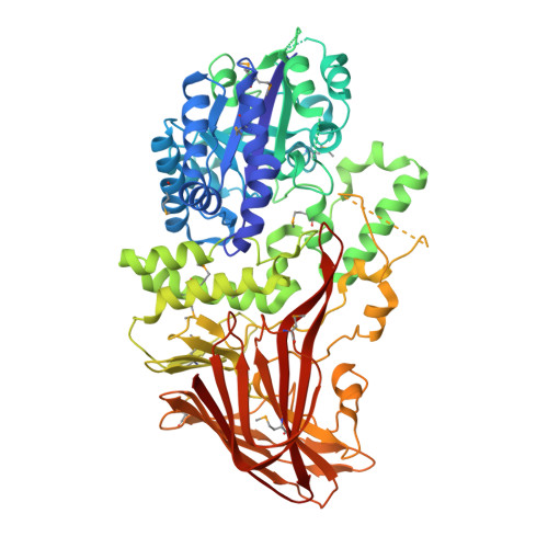
    Thermococcus litoralis 4-alpha-glucanotransferase (TLGT) belongs to glucoside hydrolase family 57 and catalyzes the disproportionation of amylose and the formation of large cyclic alpha-1,4-glucan (cycloamylose) from linear amylose. We determined the crystal structure of TLGT with and without an inhibitor, acarbose. TLGT is composed of two domains: an N-terminal domain (domain I), which contains a (beta/alpha)7 barrel fold, and a C-terminal domain (domain II), which has a twisted beta-sandwich fold. In the structure of TLGT complexed with acarbose, the inhibitor was bound at the cleft within domain I, indicating that domain I is a catalytic domain of TLGT. The acarbose-bound structure also clarified that Glu123 and Asp214 were the catalytic nucleophile and acid/base catalyst, respectively, and revealed the residues involved in substrate binding. It seemed that TLGT produces large cyclic glucans by preventing the production of small cyclic glucans by steric hindrance, which is achieved by three lids protruding into the active site cleft, as well as an extended active site cleft. Interestingly, domain I of TLGT shares some structural features with the catalytic domain of Golgi alpha-mannosidase from Drosophila melanogaster, which belongs to glucoside hydrolase family 38. Furthermore, the catalytic residue of the two enzymes is located in the same position. These observations suggest that families 57 and 38 evolved from a common ancestor.


  • Organizational Affiliation

    Department of Biotechnology, The University of Tokyo, 1-1-1 Yayoi, Bunkyo-ku, Tokyo 113-8657, Japan. himamur-ra@res.titech.ac.jp


Macromolecules
Find similar proteins by:  (by identity cutoff)  |  3D Structure
Entity ID: 1
MoleculeChains Sequence LengthOrganismDetailsImage
4-ALPHA-GLUCANOTRANSFERASE659Thermococcus litoralisMutation(s): 14 
EC: 2.4.1.25
UniProt
Find proteins for O32462 (Thermococcus litoralis (strain ATCC 51850 / DSM 5473 / JCM 8560 / NS-C))
Explore O32462 
Go to UniProtKB:  O32462
Entity Groups  
Sequence Clusters30% Identity50% Identity70% Identity90% Identity95% Identity100% Identity
UniProt GroupO32462
Sequence Annotations
Expand
  • Reference Sequence
Small Molecules
Modified Residues  1 Unique
IDChains TypeFormula2D DiagramParent
MSE
Query on MSE
A
L-PEPTIDE LINKINGC5 H11 N O2 SeMET
Experimental Data & Validation

Experimental Data

  • Method: X-RAY DIFFRACTION
  • Resolution: 2.80 Å
  • R-Value Free: 0.289 
  • R-Value Work: 0.221 
  • R-Value Observed: 0.224 
  • Space Group: P 64 2 2
Unit Cell:
Length ( Å )Angle ( ˚ )
a = 124.94α = 90
b = 124.94β = 90
c = 246.96γ = 120
Software Package:
Software NamePurpose
DENZOdata reduction
SCALEPACKdata scaling
SOLVEphasing
CNSrefinement

Structure Validation

View Full Validation Report


Ligand Structure Quality Assessment 


Entry History 

Deposition Data

Revision History  (Full details and data files)

  • Version 1.0: 2003-06-17
    Type: Initial release
  • Version 1.1: 2008-04-27
    Changes: Version format compliance
  • Version 1.2: 2011-07-13
    Changes: Version format compliance
  • Version 1.3: 2020-07-29
    Type: Remediation
    Reason: Carbohydrate remediation
    Changes: Data collection, Database references, Derived calculations, Structure summary