1J30

The crystal structure of sulerythrin, a rubrerythrin-like protein from a strictly aerobic and thermoacidiphilic archaeon


Experimental Data Snapshot

  • Method: X-RAY DIFFRACTION
  • Resolution: 1.70 Å
  • R-Value Free: 0.223 
  • R-Value Work: 0.192 
  • R-Value Observed: 0.192 

wwPDB Validation   3D Report Full Report


This is version 1.3 of the entry. See complete history


Literature

The Crystal Structure of Sulerythrin, A Rubrerythrin-like Protein from A Strictly Aerobic Archaeon, Sulfolobus tokodaii strain 7, shows unexpected domain swapping

Fushinobu, S.Shoun, H.Wakagi, T.

(2003) Biochemistry 42: 11707-11715

  • DOI: https://doi.org/10.1021/bi034220b
  • Primary Citation of Related Structures:  
    1J30

  • PubMed Abstract: 

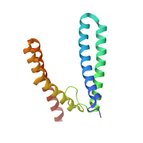
    Sulerythrin is the first rubrerythrin-like protein to be isolated from an aerobic organism, Sulfolobus tokodaii strain 7, and it lacks a C-terminal rubredoxin-like FeS(4) domain. The protein purified from Sulfolobus cells was crystallized, and the crystal structure was determined at 1.7 A resolution. The dimer of sulerythrin exhibited "domain-swapping" at the loop connecting alphaB and alphaC, hybrid four-helix bundles consisting of alphaA/B and alphaC/D being formed. The structure and atomic identity of the binuclear metal center were determined by means of anomalous scattering analysis. The site contained 1.0 mol of hexacoordinate Fe, 0.80-0.87 mol of tetracoordinate Zn, and 0.73-0.88 mol of putative O(2) per monomer. The metal ions were found at exchanged positions compared to those in the Fe/Zn-containing rubrerythrin from Desulfovibrio vulgaris. The results demonstrate that the binuclear metal center of rubrerythrin-like proteins is plastic in its ability to bind metal ions.


  • Organizational Affiliation

    Department of Biotechnology, The University of Tokyo, 1-1-1 Yayoi, Bunkyo-ku, Tokyo 113-8657, Japan. asfushi@mail.ecc.u-tokyo.ac.jp


Macromolecules
Find similar proteins by:  (by identity cutoff)  |  3D Structure
Entity ID: 1
MoleculeChains Sequence LengthOrganismDetailsImage
144aa long hypothetical rubrerythrin
A, B
144Sulfurisphaera tokodaii str. 7Mutation(s): 0 
UniProt
Find proteins for F9VPE5 (Sulfurisphaera tokodaii (strain DSM 16993 / JCM 10545 / NBRC 100140 / 7))
Explore F9VPE5 
Go to UniProtKB:  F9VPE5
Entity Groups  
Sequence Clusters30% Identity50% Identity70% Identity90% Identity95% Identity100% Identity
UniProt GroupF9VPE5
Sequence Annotations
Expand
  • Reference Sequence
Experimental Data & Validation

Experimental Data

  • Method: X-RAY DIFFRACTION
  • Resolution: 1.70 Å
  • R-Value Free: 0.223 
  • R-Value Work: 0.192 
  • R-Value Observed: 0.192 
  • Space Group: P 63
Unit Cell:
Length ( Å )Angle ( ˚ )
a = 72.428α = 90
b = 72.428β = 90
c = 98.208γ = 120
Software Package:
Software NamePurpose
CNSrefinement
MOSFLMdata reduction
CCP4data scaling
MOLREPphasing

Structure Validation

View Full Validation Report


Entry History 

Deposition Data

Revision History  (Full details and data files)

  • Version 1.0: 2003-10-14
    Type: Initial release
  • Version 1.1: 2008-04-27
    Changes: Version format compliance
  • Version 1.2: 2011-07-13
    Changes: Source and taxonomy, Version format compliance
  • Version 1.3: 2023-10-25
    Changes: Data collection, Database references, Derived calculations, Refinement description