1F1M

CRYSTAL STRUCTURE OF OUTER SURFACE PROTEIN C (OSPC)


Experimental Data Snapshot

  • Method: X-RAY DIFFRACTION
  • Resolution: 1.80 Å
  • R-Value Free: 0.235 
  • R-Value Work: 0.201 
  • R-Value Observed: 0.201 

wwPDB Validation   3D Report Full Report


This is version 1.5 of the entry. See complete history


Literature

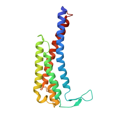
Crystal structure of outer surface protein C (OspC) from the Lyme disease spirochete, Borrelia burgdorferi.

Kumaran, D.Eswaramoorthy, S.Luft, B.J.Koide, S.Dunn, J.J.Lawson, C.L.Swaminathan, S.

(2001) EMBO J 20: 971-978

  • DOI: https://doi.org/10.1093/emboj/20.5.971
  • Primary Citation of Related Structures:  
    1F1M, 1GGQ

  • PubMed Abstract: 

    Outer surface protein C (OspC) is a major antigen on the surface of the Lyme disease spirochete, Borrelia burgdorferi, when it is being transmitted to humans. Crystal structures of OspC have been determined for strains HB19 and B31 to 1.8 and 2.5 A resolution, respectively. The three-dimensional structure is predominantly helical. This is in contrast to the structure of OspA, a major surface protein mainly present when spirochetes are residing in the midgut of unfed ticks, which is mostly beta-sheet. The surface of OspC that would project away from the spirochete's membrane has a region of strong negative electrostatic potential which may be involved in binding to positively charged host ligands. This feature is present only on OspCs from strains known to cause invasive human disease.


  • Organizational Affiliation

    Biology Department, Brookhaven National Laboratory, Upton, NY 11973, USA.


Macromolecules
Find similar proteins by:  (by identity cutoff)  |  3D Structure
Entity ID: 1
MoleculeChains Sequence LengthOrganismDetailsImage
OUTER SURFACE PROTEIN C
A, B, C, D
164Borreliella burgdorferiMutation(s): 1 
UniProt
Find proteins for Q9AGB1 (Borreliella burgdorferi)
Explore Q9AGB1 
Go to UniProtKB:  Q9AGB1
Entity Groups  
Sequence Clusters30% Identity50% Identity70% Identity90% Identity95% Identity100% Identity
UniProt GroupQ9AGB1
Sequence Annotations
Expand
  • Reference Sequence
Experimental Data & Validation

Experimental Data

  • Method: X-RAY DIFFRACTION
  • Resolution: 1.80 Å
  • R-Value Free: 0.235 
  • R-Value Work: 0.201 
  • R-Value Observed: 0.201 
  • Space Group: P 1 21 1
Unit Cell:
Length ( Å )Angle ( ˚ )
a = 66.323α = 90
b = 46.252β = 99.08
c = 111.784γ = 90
Software Package:
Software NamePurpose
PHASESphasing
SHARPphasing
CNSrefinement
MARMADdata reduction
DENZOdata reduction
SCALEPACKdata scaling

Structure Validation

View Full Validation Report


Entry History 

Deposition Data

Revision History  (Full details and data files)

  • Version 1.0: 2001-04-04
    Type: Initial release
  • Version 1.1: 2008-04-27
    Changes: Version format compliance
  • Version 1.2: 2011-07-13
    Changes: Version format compliance
  • Version 1.3: 2017-10-04
    Changes: Refinement description
  • Version 1.4: 2021-11-03
    Changes: Database references, Derived calculations
  • Version 1.5: 2024-02-07
    Changes: Data collection