7DEG

Cryo-EM structure of a heme-copper terminal oxidase dimer provides insights into its catalytic mechanism


Experimental Data Snapshot

  • Method: ELECTRON MICROSCOPY
  • Resolution: 3.40 Å
  • Aggregation State: PARTICLE 
  • Reconstruction Method: SINGLE PARTICLE 

wwPDB Validation   3D Report Full Report


This is version 1.0 of the entry. See complete history


Literature

The Unusual Homodimer of a Heme-Copper Terminal Oxidase Allows Itself to Utilize Two Electron Donors.

Zhu, G.Zeng, H.Zhang, S.Juli, J.Tai, L.Zhang, D.Pang, X.Zhang, Y.Lam, S.M.Zhu, Y.Peng, G.Michel, H.Sun, F.

(2021) Angew Chem Int Ed Engl 60: 13323-13330

  • DOI: https://doi.org/10.1002/anie.202016785
  • Primary Citation of Related Structures:  
    7DEG

  • PubMed Abstract: 

    The heme-copper oxidase superfamily comprises cytochrome c and ubiquinol oxidases. These enzymes catalyze the transfer of electrons from different electron donors onto molecular oxygen. A B-family cytochrome c oxidase from the hyperthermophilic bacterium Aquifex aeolicus was discovered previously to be able to use both cytochrome c and naphthoquinol as electron donors. Its molecular mechanism as well as the evolutionary significance are yet unknown. Here we solved its 3.4 Å resolution electron cryo-microscopic structure and discovered a novel dimeric structure mediated by subunit I (CoxA2) that would be essential for naphthoquinol binding and oxidation. The unique structural features in both proton and oxygen pathways suggest an evolutionary adaptation of this oxidase to its hyperthermophilic environment. Our results add a new conceptual understanding of structural variation of cytochrome c oxidases in different species.


  • Organizational Affiliation

    National Key Laboratory of Biomacromolecules, CAS Center for Excellence in Biomacromolecules, Institute of Biophysics, Chinese Academy of Sciences, 15 Datun Road, Chaoyang District, Beijing, 100101, China.


Macromolecules
Find similar proteins by:  (by identity cutoff)  |  3D Structure
Entity ID: 1
MoleculeChains Sequence LengthOrganismDetailsImage
Cytochrome c oxidase subunit I
A, D
587Aquifex aeolicus VF5Mutation(s): 0 
Membrane Entity: Yes 
UniProt
Find proteins for O67937 (Aquifex aeolicus (strain VF5))
Explore O67937 
Go to UniProtKB:  O67937
Entity Groups  
Sequence Clusters30% Identity50% Identity70% Identity90% Identity95% Identity100% Identity
UniProt GroupO67937
Sequence Annotations
Expand
  • Reference Sequence
Find similar proteins by:  (by identity cutoff)  |  3D Structure
Entity ID: 2
MoleculeChains Sequence LengthOrganismDetailsImage
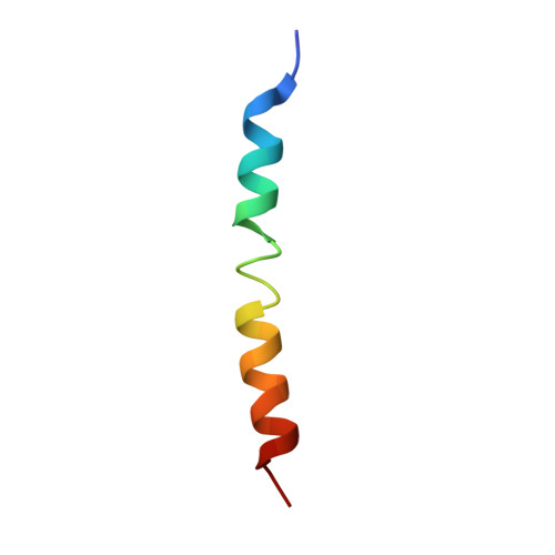
Cytochrome oxidase subunit IIaB [auth C],
E [auth F]
32Aquifex aeolicusMutation(s): 0 
Membrane Entity: Yes 
UniProt
Find proteins for G0LWX8 (Aquifex aeolicus)
Explore G0LWX8 
Go to UniProtKB:  G0LWX8
Entity Groups  
Sequence Clusters30% Identity50% Identity70% Identity90% Identity95% Identity100% Identity
UniProt GroupG0LWX8
Sequence Annotations
Expand
  • Reference Sequence
Find similar proteins by:  (by identity cutoff)  |  3D Structure
Entity ID: 3
MoleculeChains Sequence LengthOrganismDetailsImage
Cytochrome oxidase subunit IIC [auth B],
F [auth E]
147Aquifex aeolicusMutation(s): 0 
UniProt
Find proteins for G5DGC8 (Aquifex aeolicus)
Explore G5DGC8 
Go to UniProtKB:  G5DGC8
Entity Groups  
Sequence Clusters30% Identity50% Identity70% Identity90% Identity95% Identity100% Identity
UniProt GroupG5DGC8
Sequence Annotations
Expand
  • Reference Sequence
Small Molecules
Ligands 7 Unique
IDChains Name / Formula / InChI Key2D Diagram3D Interactions
HAS
Query on HAS

Download Ideal Coordinates CCD File 
G [auth A],
Q [auth D]
HEME-AS
C54 H64 Fe N4 O6
PDYODZVCODUKFH-ZOMLSHGTSA-L
PGV
Query on PGV

Download Ideal Coordinates CCD File 
K [auth A]
M [auth A]
N [auth C]
P [auth D]
U [auth D]
K [auth A],
M [auth A],
N [auth C],
P [auth D],
U [auth D],
W [auth F]
(1R)-2-{[{[(2S)-2,3-DIHYDROXYPROPYL]OXY}(HYDROXY)PHOSPHORYL]OXY}-1-[(PALMITOYLOXY)METHYL]ETHYL (11E)-OCTADEC-11-ENOATE
C40 H77 O10 P
ADYWCMPUNIVOEA-GPJPVTGXSA-N
3PE
Query on 3PE

Download Ideal Coordinates CCD File 
L [auth A],
V [auth D]
1,2-Distearoyl-sn-glycerophosphoethanolamine
C41 H82 N O8 P
LVNGJLRDBYCPGB-LDLOPFEMSA-N
DLX
Query on DLX

Download Ideal Coordinates CCD File 
J [auth A],
T [auth D]
2-[(2~{E},6~{E},10~{Z},14~{Z},18~{Z},23~{R})-3,7,11,15,19,23,27-heptamethyloctacosa-2,6,10,14,18-pentaenyl]naphthalene-1,4-dione
C45 H66 O2
HQTZEMLTZXAZEE-ZTUUOAHYSA-N
HEM
Query on HEM

Download Ideal Coordinates CCD File 
H [auth A],
R [auth D]
PROTOPORPHYRIN IX CONTAINING FE
C34 H32 Fe N4 O4
KABFMIBPWCXCRK-RGGAHWMASA-L
CUA
Query on CUA

Download Ideal Coordinates CCD File 
O [auth B],
X [auth E]
DINUCLEAR COPPER ION
Cu2
ALKZAGKDWUSJED-UHFFFAOYSA-N
CU
Query on CU

Download Ideal Coordinates CCD File 
I [auth A],
S [auth D]
COPPER (II) ION
Cu
JPVYNHNXODAKFH-UHFFFAOYSA-N
Experimental Data & Validation

Experimental Data

  • Method: ELECTRON MICROSCOPY
  • Resolution: 3.40 Å
  • Aggregation State: PARTICLE 
  • Reconstruction Method: SINGLE PARTICLE 

Structure Validation

View Full Validation Report


Entry History 

Deposition Data

Revision History  (Full details and data files)

  • Version 1.0: 2021-08-04
    Type: Initial release