6Z9C

Structure of human POLDIP2, a multifaceted adaptor protein in metabolism and genome stability


Experimental Data Snapshot

  • Method: X-RAY DIFFRACTION
  • Resolution: 2.80 Å
  • R-Value Free: 0.288 
  • R-Value Work: 0.216 
  • R-Value Observed: 0.220 

wwPDB Validation   3D Report Full Report


This is version 1.3 of the entry. See complete history


Literature

Crystal structure and molecular dynamics of human POLDIP2, a multifaceted adaptor protein in metabolism and genome stability.

Kulik, A.A.Maruszczak, K.K.Thomas, D.C.Nabi-Aldridge, N.L.A.Carr, M.Bingham, R.J.Cooper, C.D.O.

(2021) Protein Sci 30: 1196-1209

  • DOI: https://doi.org/10.1002/pro.4085
  • Primary Citation of Related Structures:  
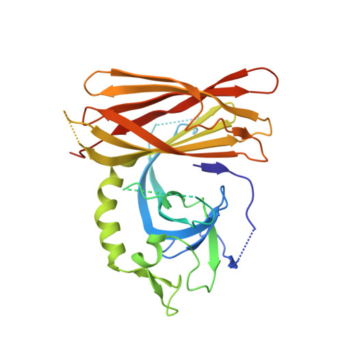
    6Z9C

  • PubMed Abstract: 

    Polymerase δ-interacting protein 2 (POLDIP2, PDIP38) is a multifaceted, "moonlighting" protein, involved in binding protein partners from many different cellular processes, including mitochondrial metabolism and DNA replication and repair. How POLDIP2 interacts with many different proteins is unknown. Towards this goal, we present the crystal structure of POLDIP2 to 2.8 Å, which exhibited a compact two-domain β-strand-rich globular structure, confirmed by circular dichroism and small angle X-ray scattering approaches. POLDIP2 comprised canonical DUF525 and YccV domains, but with a conserved domain linker packed tightly, resulting in an "extended" YccV module. A central channel was observed, which we hypothesize could influence structural changes potentially mediated by redox conditions, following observation of a modified cysteine residue in the channel. Unstructured regions were rebuilt by ab initio modelling to generate a model of full-length POLDIP2. Molecular dynamics simulations revealed a highly dynamic N-terminal region tethered to the YccV-domain by an extended linker, potentially facilitating interactions with distal binding partners. Models of POLDIP2 complexed with two of its partners, PrimPol and PCNA, indicated that dynamic flexibility of the POLDIP2 N-terminus and loop regions likely mediate protein interactions.


  • Organizational Affiliation

    Department of Biological and Geographical Sciences, School of Applied Sciences, University of Huddersfield, Huddersfield, UK.


Macromolecules
Find similar proteins by:  (by identity cutoff)  |  3D Structure
Entity ID: 1
MoleculeChains Sequence LengthOrganismDetailsImage
Polymerase delta-interacting protein 2320Homo sapiensMutation(s): 0 
Gene Names: POLDIP2PDIP38POLD4HSPC017
UniProt & NIH Common Fund Data Resources
Find proteins for Q9Y2S7 (Homo sapiens)
Explore Q9Y2S7 
Go to UniProtKB:  Q9Y2S7
PHAROS:  Q9Y2S7
GTEx:  ENSG00000004142 
Entity Groups  
Sequence Clusters30% Identity50% Identity70% Identity90% Identity95% Identity100% Identity
UniProt GroupQ9Y2S7
Sequence Annotations
Expand
  • Reference Sequence
Small Molecules
Ligands 1 Unique
IDChains Name / Formula / InChI Key2D Diagram3D Interactions
NA
Query on NA

Download Ideal Coordinates CCD File 
B [auth A]SODIUM ION
Na
FKNQFGJONOIPTF-UHFFFAOYSA-N
Experimental Data & Validation

Experimental Data

  • Method: X-RAY DIFFRACTION
  • Resolution: 2.80 Å
  • R-Value Free: 0.288 
  • R-Value Work: 0.216 
  • R-Value Observed: 0.220 
  • Space Group: P 62
Unit Cell:
Length ( Å )Angle ( ˚ )
a = 120.138α = 90
b = 120.138β = 90
c = 49.516γ = 120
Software Package:
Software NamePurpose
SCALAdata scaling
PHASERphasing
REFMACrefinement
PDB_EXTRACTdata extraction
PROTEUM2data reduction

Structure Validation

View Full Validation Report


Entry History 

Deposition Data

Revision History  (Full details and data files)

  • Version 1.0: 2020-06-17
    Type: Initial release
  • Version 1.1: 2021-05-05
    Changes: Database references
  • Version 1.2: 2021-06-02
    Changes: Database references
  • Version 1.3: 2024-01-24
    Changes: Data collection, Database references, Refinement description