6V6H

Crystal structure of histidine ammonia-lyase from Trypanosoma cruzi


Experimental Data Snapshot

  • Method: X-RAY DIFFRACTION
  • Resolution: 2.55 Å
  • R-Value Free: 0.217 
  • R-Value Work: 0.179 
  • R-Value Observed: 0.180 

wwPDB Validation   3D Report Full Report


This is version 2.0 of the entry. See complete history


Literature

Crystal structure of histidine ammonia-lyase from Trypanosoma cruzi.

Miranda, R.R.Silva, M.Barison, M.J.Silber, A.M.Iulek, J.

(2020) Biochimie 175: 181-188

  • DOI: https://doi.org/10.1016/j.biochi.2020.05.009
  • Primary Citation of Related Structures:  
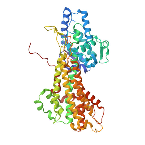
    6V6H

  • PubMed Abstract: 

    Chagas disease is one of seventeen neglected tropical diseases according to the World Health Organization (WHO). The histidine-glutamate metabolic pathway is an oxidative route that has shown to be relevant for the bioenergetics in Trypanosoma cruzi, the etiological agent for Chagas disease. Histidine ammonia-lyase participates in the first stage of the histidine catabolism, catalyzing the conversion of l-histidine into urocanate. This work presents the three-dimensional (3D) structure of Trypanosoma cruzi histidine ammonia-lyase enzyme (TcHAL) and some comparisons of it to homologous structures. The enzyme was expressed, purified and assayed for crystallization, what allowed the obtainment of crystals of sufficient quality to collect X-ray diffraction data up to 2.55 Å resolution. After refinement, some structural analyses indicated that the structure does not contain the active site protection domain, in opposition to previously known 3D structures from plants and fungi phenylalanine ammonia-lyase, therefore, it is the first structure of eukaryotic ammonia-lyases that lacks this domain.


  • Organizational Affiliation

    Department of Chemistry, State University of Ponta Grossa, Brazil.


Macromolecules
Find similar proteins by:  (by identity cutoff)  |  3D Structure
Entity ID: 1
MoleculeChains Sequence LengthOrganismDetailsImage
Histidine ammonia-lyase
A, B, C, D
538Trypanosoma cruziMutation(s): 1 
EC: 4.3.1.3
UniProt
Find proteins for Q4E133 (Trypanosoma cruzi (strain CL Brener))
Explore Q4E133 
Go to UniProtKB:  Q4E133
Entity Groups  
Sequence Clusters30% Identity50% Identity70% Identity90% Identity95% Identity100% Identity
UniProt GroupQ4E133
Sequence Annotations
Expand
  • Reference Sequence
Small Molecules
Modified Residues  2 Unique
IDChains TypeFormula2D DiagramParent
CSS
Query on CSS
A, B, C, D
L-PEPTIDE LINKINGC3 H7 N O2 S2CYS
MDO
Query on MDO
A, B, C, D
L-PEPTIDE LINKINGC8 H11 N3 O3ALA, SER, GLY
Experimental Data & Validation

Experimental Data

  • Method: X-RAY DIFFRACTION
  • Resolution: 2.55 Å
  • R-Value Free: 0.217 
  • R-Value Work: 0.179 
  • R-Value Observed: 0.180 
  • Space Group: P 21 21 21
Unit Cell:
Length ( Å )Angle ( ˚ )
a = 87.812α = 90
b = 144.555β = 90
c = 173.762γ = 90
Software Package:
Software NamePurpose
PHENIXrefinement
PDB_EXTRACTdata extraction
XDSdata reduction
PHASERphasing

Structure Validation

View Full Validation Report


Entry History & Funding Information

Deposition Data


Funding OrganizationLocationGrant Number
Brazilian National Council for Scientific and Technological DevelopmentBelize573607/2008-7 and 08/57910-0

Revision History  (Full details and data files)

  • Version 1.0: 2020-06-10
    Type: Initial release
  • Version 1.1: 2020-07-15
    Changes: Database references
  • Version 1.2: 2023-10-11
    Changes: Advisory, Data collection, Database references, Derived calculations, Refinement description
  • Version 2.0: 2023-11-15
    Changes: Atomic model, Data collection, Derived calculations