6T6N

Crystal structure of Klebsiella pneumoniae FabG2(NADH-dependent) in complex with NADH at 2.5 A resolution


Experimental Data Snapshot

  • Method: X-RAY DIFFRACTION
  • Resolution: 2.50 Å
  • R-Value Free: 0.211 
  • R-Value Work: 0.192 
  • R-Value Observed: 0.193 

wwPDB Validation   3D Report Full Report


Ligand Structure Quality Assessment 


This is version 1.2 of the entry. See complete history


Literature

A FabG inhibitor targeting an allosteric binding site inhibits several orthologs from Gram-negative ESKAPE pathogens.

Vella, P.Rudraraju, R.S.Lundback, T.Axelsson, H.Almqvist, H.Vallin, M.Schneider, G.Schnell, R.

(2021) Bioorg Med Chem 30: 115898-115898

  • DOI: https://doi.org/10.1016/j.bmc.2020.115898
  • Primary Citation of Related Structures:  
    6T5X, 6T60, 6T62, 6T65, 6T6N, 6T6P, 6T77, 6T7M

  • PubMed Abstract: 

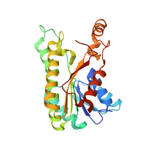
    The spread of antibiotic resistance within the ESKAPE group of human pathogenic bacteria poses severe challenges in the treatment of infections and maintenance of safe hospital environments. This motivates efforts to validate novel target proteins within these species that could be pursued as potential targets for antibiotic development. Genetic data suggest that the enzyme FabG, which is part of the bacterial fatty acid biosynthetic system FAS-II, is essential in several ESKAPE pathogens. FabG catalyzes the NADPH dependent reduction of 3-keto-acyl-ACP during fatty acid elongation, thus enabling lipid supply for production and maintenance of the cell envelope. Here we report on small-molecule screening on the FabG enzymes from A. baumannii and S. typhimurium to identify a set of µM inhibitors, with the most potent representative (1) demonstrating activity against six FabG-orthologues. A co-crystal structure with FabG from A. baumannii (PDB:6T65) confirms inhibitor binding at an allosteric site located in the subunit interface, as previously demonstrated for other sub-µM inhibitors of FabG from P. aeruginosa. We show that inhibitor binding distorts the oligomerization interface in the FabG tetramer and displaces crucial residues involved in the interaction with the co-substrate NADPH. These observations suggest a conserved allosteric site across the FabG family, which can be potentially targeted for interference with fatty acid biosynthesis in clinically relevant ESKAPE pathogens.


  • Organizational Affiliation

    Department of Medical Biochemistry and Biophysics, Karolinska Institutet, S-17 165 Stockholm, Sweden.


Macromolecules
Find similar proteins by:  (by identity cutoff)  |  3D Structure
Entity ID: 1
MoleculeChains Sequence LengthOrganismDetailsImage
3-oxoacyl-[acyl-carrier protein] reductase
A, B, C, D, E
A, B, C, D, E, F, G, H
245Klebsiella pneumoniae 30684/NJST258_2Mutation(s): 1 
Gene Names: KPNJ2_02761
EC: 1.1.1.100
UniProt
Find proteins for W8VGR4 (Klebsiella pneumoniae 30684/NJST258_2)
Explore W8VGR4 
Go to UniProtKB:  W8VGR4
Entity Groups  
Sequence Clusters30% Identity50% Identity70% Identity90% Identity95% Identity100% Identity
UniProt GroupW8VGR4
Sequence Annotations
Expand
  • Reference Sequence
Small Molecules
Ligands 3 Unique
IDChains Name / Formula / InChI Key2D Diagram3D Interactions
NAI (Subject of Investigation/LOI)
Query on NAI

Download Ideal Coordinates CCD File 
AA [auth F]
FA [auth G]
J [auth A]
JA [auth H]
M [auth B]
AA [auth F],
FA [auth G],
J [auth A],
JA [auth H],
M [auth B],
Q [auth C],
U [auth D],
X [auth E]
1,4-DIHYDRONICOTINAMIDE ADENINE DINUCLEOTIDE
C21 H29 N7 O14 P2
BOPGDPNILDQYTO-NNYOXOHSSA-N
MLT
Query on MLT

Download Ideal Coordinates CCD File 
IA [auth H]D-MALATE
C4 H6 O5
BJEPYKJPYRNKOW-UWTATZPHSA-N
GOL
Query on GOL

Download Ideal Coordinates CCD File 
BA [auth G]
CA [auth G]
DA [auth G]
EA [auth G]
GA [auth H]
BA [auth G],
CA [auth G],
DA [auth G],
EA [auth G],
GA [auth H],
HA [auth H],
I [auth A],
K [auth B],
L [auth B],
N [auth C],
O [auth C],
P [auth C],
R [auth D],
S [auth D],
T [auth D],
V [auth E],
W [auth E],
Y [auth F],
Z [auth F]
GLYCEROL
C3 H8 O3
PEDCQBHIVMGVHV-UHFFFAOYSA-N
Experimental Data & Validation

Experimental Data

  • Method: X-RAY DIFFRACTION
  • Resolution: 2.50 Å
  • R-Value Free: 0.211 
  • R-Value Work: 0.192 
  • R-Value Observed: 0.193 
  • Space Group: F 2 2 2
Unit Cell:
Length ( Å )Angle ( ˚ )
a = 161.109α = 90
b = 255.316β = 90
c = 262.312γ = 90
Software Package:
Software NamePurpose
REFMACrefinement
PDB_EXTRACTdata extraction
XDSdata reduction
Aimlessdata scaling
PHASERphasing

Structure Validation

View Full Validation Report


Ligand Structure Quality Assessment 


Entry History & Funding Information

Deposition Data


Funding OrganizationLocationGrant Number
VinnovaSweden--

Revision History  (Full details and data files)

  • Version 1.0: 2020-11-18
    Type: Initial release
  • Version 1.1: 2021-06-02
    Changes: Database references
  • Version 1.2: 2024-01-24
    Changes: Data collection, Database references, Refinement description