6S9F

Drosophila OTK, extracellular domains 3-5


Experimental Data Snapshot

  • Method: X-RAY DIFFRACTION
  • Resolution: 1.97 Å
  • R-Value Free: 0.235 
  • R-Value Work: 0.207 
  • R-Value Observed: 0.208 

wwPDB Validation   3D Report Full Report


Ligand Structure Quality Assessment 


This is version 1.3 of the entry. See complete history


Literature

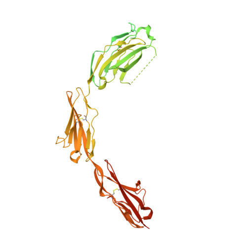
Drosophila OTK Is a Glycosaminoglycan-Binding Protein with High Conformational Flexibility.

Rozbesky, D.Monistrol, J.Jain, V.Hillier, J.Padilla-Parra, S.Jones, E.Y.

(2020) Structure 28: 507-515.e5

  • DOI: https://doi.org/10.1016/j.str.2020.02.008
  • Primary Citation of Related Structures:  
    6S9F

  • PubMed Abstract: 

    The transmembrane protein OTK plays an essential role in plexin and Wnt signaling during Drosophila development. We have determined a crystal structure of the last three domains of the OTK ectodomain and found that OTK shows high conformational flexibility resulting from mobility at the interdomain interfaces. We failed to detect direct binding between Drosophila Plexin A (PlexA) and OTK, which was suggested previously. We found that, instead of PlexA, OTK directly binds semaphorin 1a. Our binding analyses further revealed that glycosaminoglycans, heparin and heparan sulfate, are ligands for OTK and thus may play a role in the Sema1a-PlexA axon guidance system.


  • Organizational Affiliation

    Division of Structural Biology, Wellcome Centre for Human Genetics, University of Oxford, Oxford OX3 7BN, UK. Electronic address: daniel@strubi.ox.ac.uk.


Macromolecules
Find similar proteins by:  (by identity cutoff)  |  3D Structure
Entity ID: 1
MoleculeChains Sequence LengthOrganismDetailsImage
Tyrosine-protein kinase-like otk
A, B
569Drosophila melanogasterMutation(s): 0 
Gene Names: otkCG8967
UniProt
Find proteins for Q6AWJ9 (Drosophila melanogaster)
Explore Q6AWJ9 
Go to UniProtKB:  Q6AWJ9
Entity Groups  
Sequence Clusters30% Identity50% Identity70% Identity90% Identity95% Identity100% Identity
UniProt GroupQ6AWJ9
Sequence Annotations
Expand
  • Reference Sequence
Small Molecules
Ligands 2 Unique
IDChains Name / Formula / InChI Key2D Diagram3D Interactions
NAG
Query on NAG

Download Ideal Coordinates CCD File 
C [auth A]
D [auth A]
E [auth A]
F [auth A]
G [auth A]
C [auth A],
D [auth A],
E [auth A],
F [auth A],
G [auth A],
H [auth A],
M [auth B],
N [auth B],
O [auth B],
P [auth B],
Q [auth B],
R [auth B]
2-acetamido-2-deoxy-beta-D-glucopyranose
C8 H15 N O6
OVRNDRQMDRJTHS-FMDGEEDCSA-N
SO4
Query on SO4

Download Ideal Coordinates CCD File 
I [auth A]
J [auth A]
K [auth A]
L [auth A]
S [auth B]
I [auth A],
J [auth A],
K [auth A],
L [auth A],
S [auth B],
T [auth B],
U [auth B]
SULFATE ION
O4 S
QAOWNCQODCNURD-UHFFFAOYSA-L
Experimental Data & Validation

Experimental Data

  • Method: X-RAY DIFFRACTION
  • Resolution: 1.97 Å
  • R-Value Free: 0.235 
  • R-Value Work: 0.207 
  • R-Value Observed: 0.208 
  • Space Group: C 2 2 21
Unit Cell:
Length ( Å )Angle ( ˚ )
a = 80.975α = 90
b = 189.909β = 90
c = 131.906γ = 90
Software Package:
Software NamePurpose
Aimlessdata scaling
PHENIXrefinement
PDB_EXTRACTdata extraction
xia2data reduction
PHASERphasing

Structure Validation

View Full Validation Report


Ligand Structure Quality Assessment 


Entry History & Funding Information

Deposition Data


Funding OrganizationLocationGrant Number
Cancer Research UKUnited KingdomC375/A17721
Medical Research Council (United Kingdom)United KingdomMR/M000141/1

Revision History  (Full details and data files)

  • Version 1.0: 2020-03-25
    Type: Initial release
  • Version 1.1: 2020-05-13
    Changes: Database references
  • Version 1.2: 2020-07-29
    Type: Remediation
    Reason: Carbohydrate remediation
    Changes: Data collection, Derived calculations, Structure summary
  • Version 1.3: 2024-01-24
    Changes: Data collection, Database references, Refinement description, Structure summary