6QKZ

Full length GluA1/2-gamma8 complex


Experimental Data Snapshot

  • Method: ELECTRON MICROSCOPY
  • Resolution: 6.30 Å
  • Aggregation State: PARTICLE 
  • Reconstruction Method: SINGLE PARTICLE 

wwPDB Validation   3D Report Full Report


This is version 2.0 of the entry. See complete history


Literature

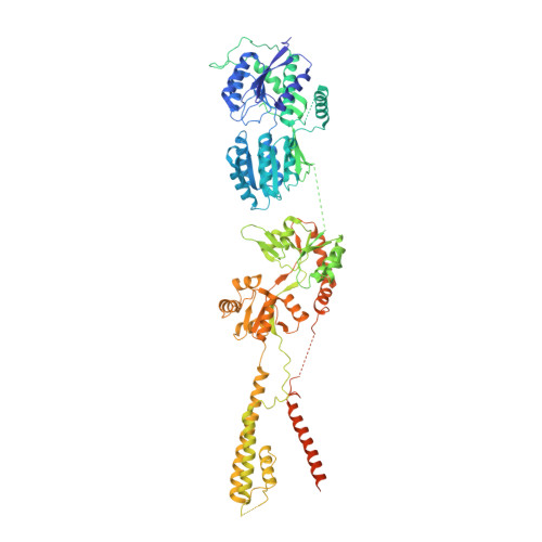
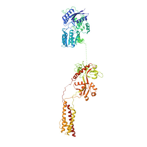
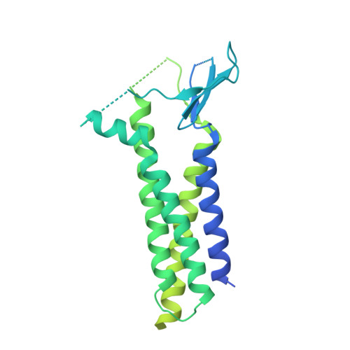
Architecture of the heteromeric GluA1/2 AMPA receptor in complex with the auxiliary subunit TARP gamma 8.

Herguedas, B.Watson, J.F.Ho, H.Cais, O.Garcia-Nafria, J.Greger, I.H.

(2019) Science 364

  • DOI: https://doi.org/10.1126/science.aav9011
  • Primary Citation of Related Structures:  
    6QKC, 6QKZ

  • PubMed Abstract: 

    AMPA-type glutamate receptors (AMPARs) mediate excitatory neurotransmission and are central regulators of synaptic plasticity, a molecular mechanism underlying learning and memory. Although AMPARs act predominantly as heteromers, structural studies have focused on homomeric assemblies. Here, we present a cryo-electron microscopy structure of the heteromeric GluA1/2 receptor associated with two transmembrane AMPAR regulatory protein (TARP) γ8 auxiliary subunits, the principal AMPAR complex at hippocampal synapses. Within the receptor, the core subunits arrange to give the GluA2 subunit dominant control of gating. This structure reveals the geometry of the Q/R site that controls calcium flux, suggests association of TARP-stabilized lipids, and demonstrates that the extracellular loop of γ8 modulates gating by selectively interacting with the GluA2 ligand-binding domain. Collectively, this structure provides a blueprint for deciphering the signal transduction mechanisms of synaptic AMPARs.


  • Organizational Affiliation

    Neurobiology Division, Medical Research Council (MRC) Laboratory of Molecular Biology, Cambridge, UK. ig@mrc-lmb.cam.ac.uk hergueda@mrc-lmb.cam.ac.uk.


Macromolecules
Find similar proteins by:  (by identity cutoff)  |  3D Structure
Entity ID: 1
MoleculeChains Sequence LengthOrganismDetailsImage
GluA1
A, C
897Rattus norvegicusMutation(s): 0 
Membrane Entity: Yes 
UniProt
Find proteins for P19490 (Rattus norvegicus)
Explore P19490 
Go to UniProtKB:  P19490
Entity Groups  
Sequence Clusters30% Identity50% Identity70% Identity90% Identity95% Identity100% Identity
UniProt GroupP19490
Sequence Annotations
Expand
  • Reference Sequence
Find similar proteins by:  (by identity cutoff)  |  3D Structure
Entity ID: 2
MoleculeChains Sequence LengthOrganismDetailsImage
Glutamate receptor 2
B, D
839Rattus norvegicusMutation(s): 0 
Gene Names: Gria2Glur2
Membrane Entity: Yes 
UniProt
Find proteins for P19491 (Rattus norvegicus)
Explore P19491 
Go to UniProtKB:  P19491
Entity Groups  
Sequence Clusters30% Identity50% Identity70% Identity90% Identity95% Identity100% Identity
UniProt GroupP19491
Sequence Annotations
Expand
  • Reference Sequence
Find similar proteins by:  (by identity cutoff)  |  3D Structure
Entity ID: 3
MoleculeChains Sequence LengthOrganismDetailsImage
Voltage-dependent calcium channel gamma-8 subunitE [auth I],
F [auth J]
423Rattus norvegicusMutation(s): 0 
Gene Names: Cacng8
Membrane Entity: Yes 
UniProt
Find proteins for Q8VHW5 (Rattus norvegicus)
Explore Q8VHW5 
Go to UniProtKB:  Q8VHW5
Entity Groups  
Sequence Clusters30% Identity50% Identity70% Identity90% Identity95% Identity100% Identity
UniProt GroupQ8VHW5
Sequence Annotations
Expand
  • Reference Sequence
Oligosaccharides

Help

Entity ID: 4
MoleculeChains Length2D Diagram Glycosylation3D Interactions
beta-D-mannopyranose-(1-4)-2-acetamido-2-deoxy-beta-D-glucopyranose-(1-4)-2-acetamido-2-deoxy-beta-D-glucopyranoseG [auth E],
K
3N-Glycosylation
Glycosylation Resources
GlyTouCan:  G15407YE
GlyCosmos:  G15407YE
GlyGen:  G15407YE
Entity ID: 5
MoleculeChains Length2D Diagram Glycosylation3D Interactions
2-acetamido-2-deoxy-beta-D-glucopyranose-(1-4)-2-acetamido-2-deoxy-beta-D-glucopyranose
H [auth F],
I [auth G],
J [auth H],
L,
M,
H [auth F],
I [auth G],
J [auth H],
L,
M,
N
2N-Glycosylation
Glycosylation Resources
GlyTouCan:  G42666HT
GlyCosmos:  G42666HT
GlyGen:  G42666HT
Small Molecules
Binding Affinity Annotations 
IDSourceBinding Affinity
E2Q BindingDB:  6QKZ Ki: min: 210, max: 1600 (nM) from 3 assay(s)
IC50: min: 2500, max: 6000 (nM) from 2 assay(s)
Experimental Data & Validation

Experimental Data

  • Method: ELECTRON MICROSCOPY
  • Resolution: 6.30 Å
  • Aggregation State: PARTICLE 
  • Reconstruction Method: SINGLE PARTICLE 
EM Software:
TaskSoftware PackageVersion
RECONSTRUCTIONRELION3.0

Structure Validation

View Full Validation Report


Entry History 

Deposition Data

Revision History  (Full details and data files)

  • Version 1.0: 2019-03-27
    Type: Initial release
  • Version 1.1: 2019-05-08
    Changes: Data collection, Database references
  • Version 1.2: 2019-11-06
    Changes: Data collection, Database references
  • Version 2.0: 2020-07-29
    Type: Remediation
    Reason: Carbohydrate remediation
    Changes: Advisory, Atomic model, Data collection, Derived calculations, Structure summary