6PX4

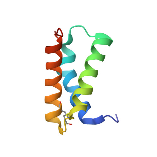
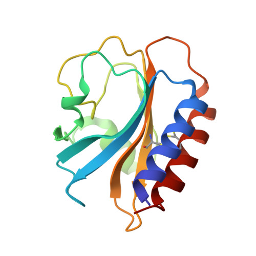
Crystal structure of the complex between periplasmic domains of antiholin RI and holin T from T4 phage, in H32


Experimental Data Snapshot

  • Method: X-RAY DIFFRACTION
  • Resolution: 1.65 Å
  • R-Value Free: 0.225 
  • R-Value Work: 0.194 
  • R-Value Observed: 0.195 

wwPDB Validation   3D Report Full Report


This is version 1.2 of the entry. See complete history


Literature

The Structural Basis of T4 Phage Lysis Control: DNA as the Signal for Lysis Inhibition.

Krieger, I.V.Kuznetsov, V.Chang, J.Y.Zhang, J.Moussa, S.H.Young, R.F.Sacchettini, J.C.

(2020) J Mol Biol 432: 4623-4636

  • DOI: https://doi.org/10.1016/j.jmb.2020.06.013
  • Primary Citation of Related Structures:  
    6PSH, 6PSK, 6PX4, 6PXE

  • PubMed Abstract: 

    Optimal phage propagation depends on the regulation of the lysis of the infected host cell. In T4 phage infection, lysis occurs when the holin protein (T) forms lesions in the host membrane. However, the lethal function of T can be blocked by an antiholin (RI) during lysis inhibition (LIN). LIN sets if the infected cell undergoes superinfection, then the lysis is delayed until host/phage ratio becomes more favorable for the release of progeny. It has been thought that a signal derived from the superinfection is required to activate RI. Here we report structures that suggest a radically different model in which RI binds to T irrespective of superinfection, causing it to accumulate in a membrane as heterotetrameric 2RI-2T complex. Moreover, we show the complex binds non-specifically to DNA, suggesting that the gDNA from the superinfecting phage serves as the LIN signal and that stabilization of the complex by DNA binding is what defines LIN. Finally, we show that soluble domain of free RI crystallizes in a domain-swapped homotetramer, which likely works as a sink for RI molecules released from the RI-T complex to ensure efficient lysis. These results constitute the first structural basis and a new model not only for the historic LIN phenomenon but also for the temporal regulation of phage lysis in general.


  • Organizational Affiliation

    Department of Biochemistry and Biophysics, Texas A&M University, College Station, TX 77843, USA.


Macromolecules
Find similar proteins by:  (by identity cutoff)  |  3D Structure
Entity ID: 1
MoleculeChains Sequence LengthOrganismDetailsImage
AntiholinA [auth R],
C [auth A]
74Escherichia phage ECML-134Mutation(s): 0 
Gene Names: ECML134_104
UniProt
Find proteins for P13304 (Enterobacteria phage T4)
Explore P13304 
Go to UniProtKB:  P13304
Entity Groups  
Sequence Clusters30% Identity50% Identity70% Identity90% Identity95% Identity100% Identity
UniProt GroupP13304
Sequence Annotations
Expand
  • Reference Sequence
Find similar proteins by:  (by identity cutoff)  |  3D Structure
Entity ID: 2
MoleculeChains Sequence LengthOrganismDetailsImage
HolinB [auth T],
D [auth B]
142Escherichia phage vB_EcoM_NBG2Mutation(s): 0 
Gene Names: vBEcoMNBG2_239
UniProt
Find proteins for P06808 (Enterobacteria phage T4)
Explore P06808 
Go to UniProtKB:  P06808
Entity Groups  
Sequence Clusters30% Identity50% Identity70% Identity90% Identity95% Identity100% Identity
UniProt GroupP06808
Sequence Annotations
Expand
  • Reference Sequence
Experimental Data & Validation

Experimental Data

  • Method: X-RAY DIFFRACTION
  • Resolution: 1.65 Å
  • R-Value Free: 0.225 
  • R-Value Work: 0.194 
  • R-Value Observed: 0.195 
  • Space Group: H 3 2
Unit Cell:
Length ( Å )Angle ( ˚ )
a = 116.546α = 90
b = 116.546β = 90
c = 223.384γ = 120
Software Package:
Software NamePurpose
PHENIXrefinement
SAINTdata reduction
SAINTdata scaling
MOLREPphasing
PDB_EXTRACTdata extraction

Structure Validation

View Full Validation Report


Entry History & Funding Information

Deposition Data


Funding OrganizationLocationGrant Number
Welch FoundationUnited StatesA-0015

Revision History  (Full details and data files)

  • Version 1.0: 2020-06-24
    Type: Initial release
  • Version 1.1: 2020-07-01
    Changes: Database references
  • Version 1.2: 2020-08-12
    Changes: Database references