6NQ3

Crystal Structure of a SUZ12-RBBP4-PHF19-JARID2 Heterotetrameric Complex


Experimental Data Snapshot

  • Method: X-RAY DIFFRACTION
  • Resolution: 2.89 Å
  • R-Value Free: 0.230 
  • R-Value Work: 0.170 
  • R-Value Observed: 0.173 

wwPDB Validation   3D Report Full Report


This is version 1.3 of the entry. See complete history


Literature

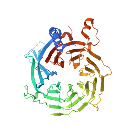
A Dimeric Structural Scaffold for PRC2-PCL Targeting to CpG Island Chromatin.

Chen, S.Jiao, L.Liu, X.Yang, X.Liu, X.

(2020) Mol Cell 77: 1265

  • DOI: https://doi.org/10.1016/j.molcel.2019.12.019
  • Primary Citation of Related Structures:  
    6NQ3

  • PubMed Abstract: 

    Diverse accessory subunits are involved in the recruitment of polycomb repressive complex 2 (PRC2) to CpG island (CGI) chromatin. Here we report the crystal structure of a SUZ12-RBBP4 complex bound to fragments of the accessory subunits PHF19 and JARID2. Unexpectedly, this complex adopts a dimeric structural architecture, accounting for PRC2 self-association that has long been implicated. The intrinsic PRC2 dimer is formed via domain swapping involving RBBP4 and the unique C2 domain of SUZ12. MTF2 and PHF19 associate with PRC2 at around the dimer interface and stabilize the dimer. Conversely, AEBP2 binding results in a drastic movement of the C2 domain, disrupting the intrinsic PRC2 dimer. PRC2 dimerization enhances CGI DNA binding by PCLs in pairs in vitro, reminiscent of the widespread phenomenon of transcription factor dimerization in active transcription. Loss of PRC2 dimerization impairs histone H3K27 trimethylation (H3K27me3) on chromatin at developmental gene loci in mouse embryonic stem cells.


  • Organizational Affiliation

    Cecil H. and Ida Green Center for Reproductive Biology Sciences and Division of Basic Research, Department of Obstetrics and Gynecology, UT Southwestern Medical Center, Dallas, TX 75390, USA; Department of Biophysics, UT Southwestern Medical Center, Dallas, TX 75390, USA.


Macromolecules
Find similar proteins by:  (by identity cutoff)  |  3D Structure
Entity ID: 1
MoleculeChains Sequence LengthOrganismDetailsImage
Histone-binding protein RBBP4
A, E
439Homo sapiensMutation(s): 0 
Gene Names: RBBP4RBAP48
UniProt & NIH Common Fund Data Resources
Find proteins for Q09028 (Homo sapiens)
Explore Q09028 
Go to UniProtKB:  Q09028
PHAROS:  Q09028
GTEx:  ENSG00000162521 
Entity Groups  
Sequence Clusters30% Identity50% Identity70% Identity90% Identity95% Identity100% Identity
UniProt GroupQ09028
Sequence Annotations
Expand
  • Reference Sequence
Find similar proteins by:  (by identity cutoff)  |  3D Structure
Entity ID: 2
MoleculeChains Sequence LengthOrganismDetailsImage
Polycomb protein SUZ12
B, F
478Homo sapiensMutation(s): 0 
Gene Names: SUZ12CHET9JJAZ1KIAA0160
UniProt & NIH Common Fund Data Resources
Find proteins for Q15022 (Homo sapiens)
Explore Q15022 
Go to UniProtKB:  Q15022
PHAROS:  Q15022
GTEx:  ENSG00000178691 
Entity Groups  
Sequence Clusters30% Identity50% Identity70% Identity90% Identity95% Identity100% Identity
UniProt GroupQ15022
Sequence Annotations
Expand
  • Reference Sequence
Find similar proteins by:  (by identity cutoff)  |  3D Structure
Entity ID: 3
MoleculeChains Sequence LengthOrganismDetailsImage
PHD finger protein 19
C, G
84Homo sapiensMutation(s): 0 
Gene Names: PHF19PCL3
UniProt & NIH Common Fund Data Resources
Find proteins for Q5T6S3 (Homo sapiens)
Explore Q5T6S3 
Go to UniProtKB:  Q5T6S3
PHAROS:  Q5T6S3
GTEx:  ENSG00000119403 
Entity Groups  
Sequence Clusters30% Identity50% Identity70% Identity90% Identity95% Identity100% Identity
UniProt GroupQ5T6S3
Sequence Annotations
Expand
  • Reference Sequence

Find similar proteins by:  Sequence   |   3D Structure  

Entity ID: 4
MoleculeChains Sequence LengthOrganismDetailsImage
Protein Jumonji
D, H
19Homo sapiensMutation(s): 0 
UniProt & NIH Common Fund Data Resources
Find proteins for Q92833 (Homo sapiens)
Explore Q92833 
Go to UniProtKB:  Q92833
PHAROS:  Q92833
GTEx:  ENSG00000008083 
Entity Groups  
Sequence Clusters30% Identity50% Identity70% Identity90% Identity95% Identity100% Identity
UniProt GroupQ92833
Sequence Annotations
Expand
  • Reference Sequence
Experimental Data & Validation

Experimental Data

  • Method: X-RAY DIFFRACTION
  • Resolution: 2.89 Å
  • R-Value Free: 0.230 
  • R-Value Work: 0.170 
  • R-Value Observed: 0.173 
  • Space Group: C 2 2 21
Unit Cell:
Length ( Å )Angle ( ˚ )
a = 127.71α = 90
b = 139.6β = 90
c = 268.08γ = 90
Software Package:
Software NamePurpose
BUSTERrefinement
HKL-2000data reduction
HKL-2000data scaling
PHASERphasing

Structure Validation

View Full Validation Report


Entry History 

Deposition Data

Revision History  (Full details and data files)

  • Version 1.0: 2020-01-29
    Type: Initial release
  • Version 1.1: 2020-02-12
    Changes: Structure summary
  • Version 1.2: 2020-04-08
    Changes: Database references
  • Version 1.3: 2024-03-13
    Changes: Data collection, Database references