6LOS

Crystal structure of mouse PEDF in complex with heterotrimeric collagen model peptide.


Experimental Data Snapshot

  • Method: X-RAY DIFFRACTION
  • Resolution: 2.48 Å
  • R-Value Free: 0.282 
  • R-Value Work: 0.223 
  • R-Value Observed: 0.228 

wwPDB Validation   3D Report Full Report


This is version 1.1 of the entry. See complete history


Literature

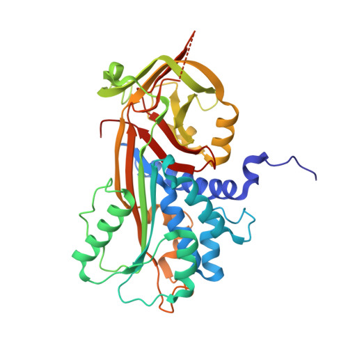
Spatiotemporal regulation of PEDF signaling by type I collagen remodeling.

Kawahara, K.Yoshida, T.Maruno, T.Oki, H.Ohkubo, T.Koide, T.Kobayashi, Y.

(2020) Proc Natl Acad Sci U S A 117: 11450-11458

  • DOI: https://doi.org/10.1073/pnas.2004034117
  • Primary Citation of Related Structures:  
    6LOS

  • PubMed Abstract: 

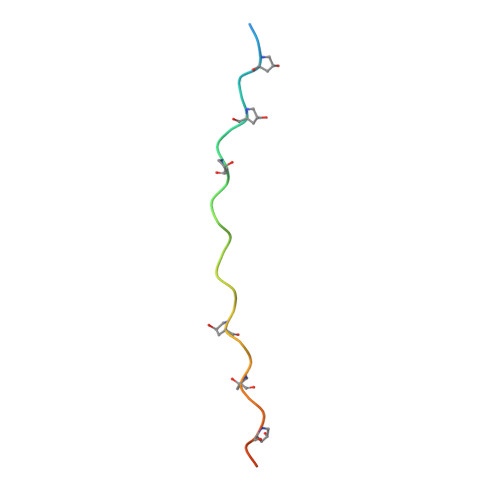
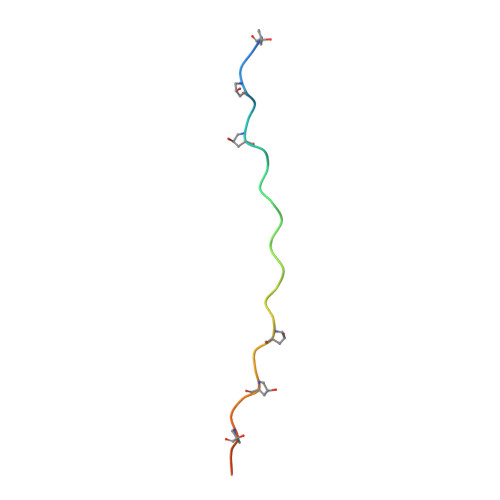
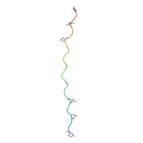
    Dynamic remodeling of the extracellular matrix affects many cellular processes, either directly or indirectly, through the regulation of soluble ligands; however, the mechanistic details of this process remain largely unknown. Here we propose that type I collagen remodeling regulates the receptor-binding activity of pigment epithelium-derived factor (PEDF), a widely expressed secreted glycoprotein that has multiple important biological functions in tissue and organ homeostasis. We determined the crystal structure of PEDF in complex with a disulfide cross-linked heterotrimeric collagen peptide, in which the α(I) chain segments-each containing the respective PEDF-binding region (residues 930 to 938)-are assembled with an α2α1α1 staggered configuration. The complex structure revealed that PEDF specifically interacts with a unique amphiphilic sequence, KGHRGFSGL, of the type I collagen α1 chain, with its proposed receptor-binding sites buried extensively. Molecular docking demonstrated that the PEDF-binding surface of type I collagen contains the cross-link-susceptible Lys930 residue of the α1 chain and provides a good foothold for stable docking with the α1(I) N-telopeptide of an adjacent triple helix in the fibril. Therefore, the binding surface is completely inaccessible if intermolecular crosslinking between two crosslink-susceptible lysyl residues, Lys9 in the N-telopeptide and Lys930, is present. These structural analyses demonstrate that PEDF molecules, once sequestered around newly synthesized pericellular collagen fibrils, are gradually liberated as collagen crosslinking increases, making them accessible for interaction with their target cell surface receptors in a spatiotemporally regulated manner.


  • Organizational Affiliation

    Graduate School of Pharmaceutical Sciences, Osaka University, Suita, 565-0871 Osaka, Japan.


Macromolecules
Find similar proteins by:  (by identity cutoff)  |  3D Structure
Entity ID: 1
MoleculeChains Sequence LengthOrganismDetailsImage
Pigment epithelium-derived factor377Mus musculusMutation(s): 0 
Gene Names: Serpinf1PedfSdf3
UniProt & NIH Common Fund Data Resources
Find proteins for P97298 (Mus musculus)
Explore P97298 
Go to UniProtKB:  P97298
IMPC:  MGI:108080
Entity Groups  
Sequence Clusters30% Identity50% Identity70% Identity90% Identity95% Identity100% Identity
UniProt GroupP97298
Sequence Annotations
Expand
  • Reference Sequence
Find similar proteins by:  (by identity cutoff)  |  3D Structure
Entity ID: 2
MoleculeChains Sequence LengthOrganismDetailsImage
Collagen model peptide, type I, alpha 2B [auth F]35Homo sapiensMutation(s): 0 
Entity Groups  
Sequence Clusters30% Identity50% Identity70% Identity90% Identity95% Identity100% Identity
Sequence Annotations
Expand
  • Reference Sequence
Find similar proteins by:  (by identity cutoff)  |  3D Structure
Entity ID: 3
MoleculeChains Sequence LengthOrganismDetailsImage
Collagen model peptide, type I, alpha 1C [auth G]34Homo sapiensMutation(s): 0 
Entity Groups  
Sequence Clusters30% Identity50% Identity70% Identity90% Identity95% Identity100% Identity
Sequence Annotations
Expand
  • Reference Sequence
Find similar proteins by:  (by identity cutoff)  |  3D Structure
Entity ID: 4
MoleculeChains Sequence LengthOrganismDetailsImage
Collagen model peptide, type I, alpha 1D [auth H]33Homo sapiensMutation(s): 0 
Entity Groups  
Sequence Clusters30% Identity50% Identity70% Identity90% Identity95% Identity100% Identity
Sequence Annotations
Expand
  • Reference Sequence
Small Molecules
Modified Residues  1 Unique
IDChains TypeFormula2D DiagramParent
HYP
Query on HYP
B [auth F]L-PEPTIDE LINKINGC5 H9 N O3PRO
Experimental Data & Validation

Experimental Data

  • Method: X-RAY DIFFRACTION
  • Resolution: 2.48 Å
  • R-Value Free: 0.282 
  • R-Value Work: 0.223 
  • R-Value Observed: 0.228 
  • Space Group: C 2 2 21
Unit Cell:
Length ( Å )Angle ( ˚ )
a = 95.378α = 90
b = 147.773β = 90
c = 91.548γ = 90
Software Package:
Software NamePurpose
PHENIXrefinement
HKL-2000data scaling
PDB_EXTRACTdata extraction
PHENIXphasing
HKL-2000data reduction

Structure Validation

View Full Validation Report


Entry History 

Deposition Data

Revision History  (Full details and data files)

  • Version 1.0: 2020-09-02
    Type: Initial release
  • Version 1.1: 2023-11-29
    Changes: Data collection, Database references, Refinement description