6KU0

Crystal structure of MyoVa-GTD in complex with MICAL1-GTBM


Experimental Data Snapshot

  • Method: X-RAY DIFFRACTION
  • Resolution: 1.60 Å
  • R-Value Free: 0.199 
  • R-Value Work: 0.161 
  • R-Value Observed: 0.162 

wwPDB Validation   3D Report Full Report


This is version 1.2 of the entry. See complete history


Literature

F-actin disassembly factor MICAL1 binding to Myosin Va mediates cargo unloading during cytokinesis.

Niu, F.Sun, K.Wei, W.Yu, C.Wei, Z.

(2020) Sci Adv 6

  • DOI: https://doi.org/10.1126/sciadv.abb1307
  • Primary Citation of Related Structures:  
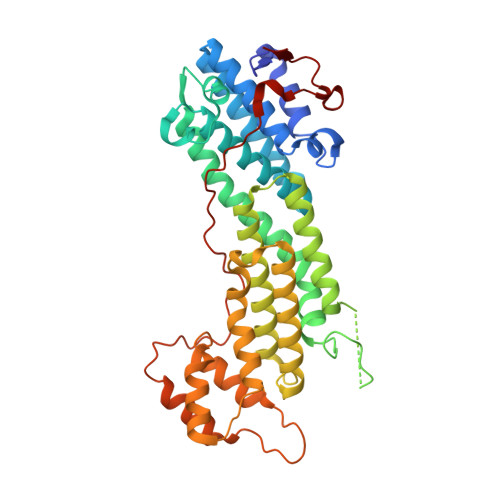
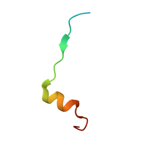
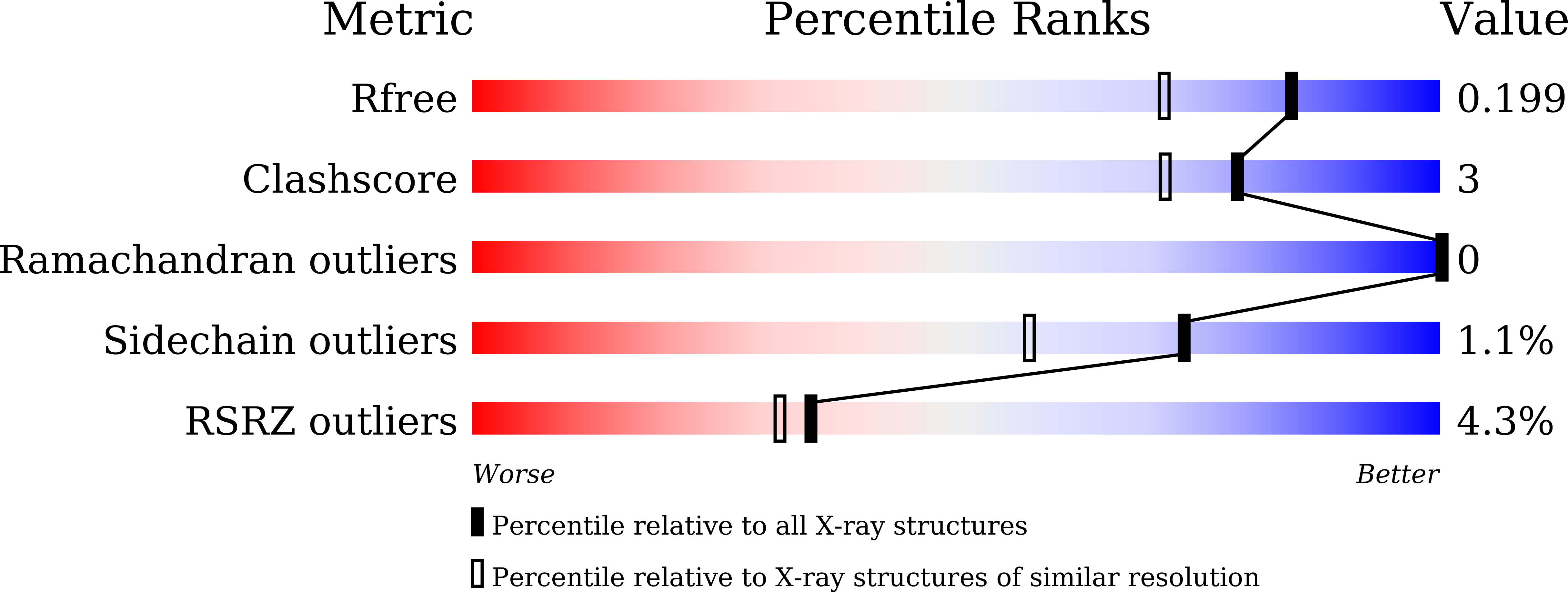
    6KU0

  • PubMed Abstract: 

    Motor-mediated intracellular trafficking requires motors to position cargoes at proper locations. Myosin Va (MyoVa), an actin-based motor, is a classic model for studying cargo transport. However, the molecular basis underlying cargo unloading in MyoVa-mediated transport has remained enigmatic. We have identified MICAL1, an F-actin disassembly regulator, as a binding partner of MyoVa and shown that MICAL1-MyoVa interaction is critical for localization of MyoVa at the midbody. By binding to MICAL1, MyoVa-mediated transport is terminated, resulting in vesicle unloading at the midbody for efficient cytokinesis. The MyoVa/MICAL1 complex structure reveals that MICAL1 and F-actin assembly factors, Spires, share an overlapped binding surface on MyoVa, suggesting a regulatory role of F-actin dynamics in cargo unloading. Down-regulating F-actin disassembly by a MICAL1 mutant significantly reduces MyoVa and vesicles accumulating at the midbody. Collectively, our findings demonstrate that MyoVa binds to MICAL1 at the midbody destination and triggers F-actin disassembly to unload the vesicle cargo.


  • Organizational Affiliation

    Department of Biology, Southern University of Science and Technology, Shenzhen, Guangdong, China.


Macromolecules
Find similar proteins by:  (by identity cutoff)  |  3D Structure
Entity ID: 1
MoleculeChains Sequence LengthOrganismDetailsImage
Unconventional myosin-Va
A, C
389Mus musculusMutation(s): 0 
Gene Names: Myo5aDilute
UniProt & NIH Common Fund Data Resources
Find proteins for Q99104 (Mus musculus)
Explore Q99104 
Go to UniProtKB:  Q99104
IMPC:  MGI:105976
Entity Groups  
Sequence Clusters30% Identity50% Identity70% Identity90% Identity95% Identity100% Identity
UniProt GroupQ99104
Sequence Annotations
Expand
  • Reference Sequence
Find similar proteins by:  (by identity cutoff)  |  3D Structure
Entity ID: 2
MoleculeChains Sequence LengthOrganismDetailsImage
Peptide from [F-actin]-monooxygenase MICAL1
B, D
27Homo sapiensMutation(s): 0 
Gene Names: MICAL1MICALNICAL
UniProt & NIH Common Fund Data Resources
Find proteins for Q8TDZ2 (Homo sapiens)
Explore Q8TDZ2 
Go to UniProtKB:  Q8TDZ2
PHAROS:  Q8TDZ2
GTEx:  ENSG00000135596 
Entity Groups  
Sequence Clusters30% Identity50% Identity70% Identity90% Identity95% Identity100% Identity
UniProt GroupQ8TDZ2
Sequence Annotations
Expand
  • Reference Sequence
Experimental Data & Validation

Experimental Data

  • Method: X-RAY DIFFRACTION
  • Resolution: 1.60 Å
  • R-Value Free: 0.199 
  • R-Value Work: 0.161 
  • R-Value Observed: 0.162 
  • Space Group: P 1
Unit Cell:
Length ( Å )Angle ( ˚ )
a = 60.293α = 65.78
b = 63.22β = 86.71
c = 74.418γ = 85.81
Software Package:
Software NamePurpose
PHENIXrefinement
HKL-2000data scaling
PDB_EXTRACTdata extraction
HKL-2000data reduction
PHASERphasing

Structure Validation

View Full Validation Report


Entry History & Funding Information

Deposition Data


Funding OrganizationLocationGrant Number
National Natural Science Foundation of ChinaChina31800643
National Natural Science Foundation of ChinaChina31770791
National Natural Science Foundation of ChinaChina31570741

Revision History  (Full details and data files)

  • Version 1.0: 2020-09-02
    Type: Initial release
  • Version 1.1: 2020-11-18
    Changes: Database references
  • Version 1.2: 2023-11-22
    Changes: Data collection, Database references, Refinement description