6KG7

Cryo-EM Structure of the Mammalian Tactile Channel Piezo2


Experimental Data Snapshot

  • Method: ELECTRON MICROSCOPY
  • Resolution: 3.80 Å
  • Aggregation State: PARTICLE 
  • Reconstruction Method: SINGLE PARTICLE 

wwPDB Validation   3D Report Full Report


This is version 1.3 of the entry. See complete history


Literature

Structure and mechanogating of the mammalian tactile channel PIEZO2.

Wang, L.Zhou, H.Zhang, M.Liu, W.Deng, T.Zhao, Q.Li, Y.Lei, J.Li, X.Xiao, B.

(2019) Nature 573: 225-229

  • DOI: https://doi.org/10.1038/s41586-019-1505-8
  • Primary Citation of Related Structures:  
    6KG7

  • PubMed Abstract: 

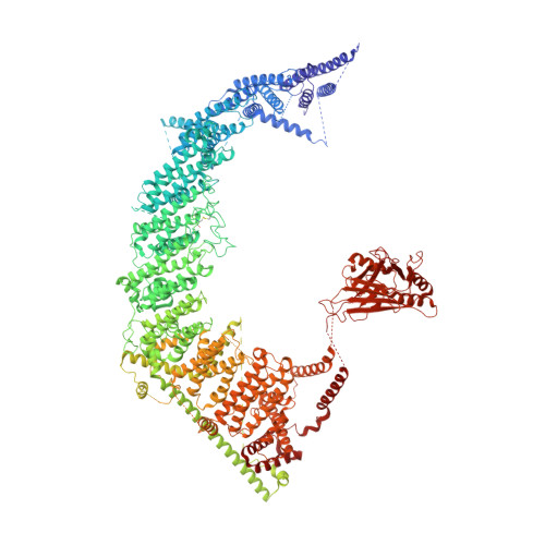
    PIEZO2 is a mechanosensitive cation channel that has a key role in sensing touch, tactile pain, breathing and blood pressure. Here we describe the cryo-electron microscopy structure of mouse PIEZO2, which is a three-bladed, propeller-like trimer that comprises 114 transmembrane helices (38 per protomer). Transmembrane helices 1-36 (TM1-36) are folded into nine tandem units of four transmembrane helices each to form the unusual non-planar blades. The three blades are collectively curved into a nano-dome of 28-nm diameter and 10-nm depth, with an extracellular cap-like structure embedded in the centre and a 9-nm-long intracellular beam connecting to the central pore. TM38 and the C-terminal domain are surrounded by the anchor domain and TM37, and enclose the central pore with both transmembrane and cytoplasmic constriction sites. Structural comparison between PIEZO2 and its homologue PIEZO1 reveals that the transmembrane constriction site might act as a transmembrane gate that is controlled by the cap domain. Together, our studies provide insights into the structure and mechanogating mechanism of Piezo channels.


  • Organizational Affiliation

    State Key Laboratory of Membrane Biology, Beijing Advanced Innovation Center for Structural Biology, Tsinghua-Peking Joint Center for Life Sciences, IDG/McGovern Institute for Brain Research, School of Pharmaceutical Sciences, Tsinghua University, Beijing, China.


Macromolecules
Find similar proteins by:  (by identity cutoff)  |  3D Structure
Entity ID: 1
MoleculeChains Sequence LengthOrganismDetailsImage
Piezo-type mechanosensitive ion channel component 2
A, B, C
2,822Mus musculusMutation(s): 0 
Gene Names: Piezo2Fam38b
Membrane Entity: Yes 
UniProt & NIH Common Fund Data Resources
Find proteins for Q8CD54 (Mus musculus)
Explore Q8CD54 
Go to UniProtKB:  Q8CD54
IMPC:  MGI:1918781
Entity Groups  
Sequence Clusters30% Identity50% Identity70% Identity90% Identity95% Identity100% Identity
UniProt GroupQ8CD54
Sequence Annotations
Expand
  • Reference Sequence
Small Molecules
Ligands 1 Unique
IDChains Name / Formula / InChI Key2D Diagram3D Interactions
NAG
Query on NAG

Download Ideal Coordinates CCD File 
D [auth A]
E [auth A]
F [auth A]
G [auth A]
H [auth B]
D [auth A],
E [auth A],
F [auth A],
G [auth A],
H [auth B],
I [auth B],
J [auth B],
K [auth B],
L [auth C],
M [auth C],
N [auth C],
O [auth C]
2-acetamido-2-deoxy-beta-D-glucopyranose
C8 H15 N O6
OVRNDRQMDRJTHS-FMDGEEDCSA-N
Experimental Data & Validation

Experimental Data

  • Method: ELECTRON MICROSCOPY
  • Resolution: 3.80 Å
  • Aggregation State: PARTICLE 
  • Reconstruction Method: SINGLE PARTICLE 
EM Software:
TaskSoftware PackageVersion
RECONSTRUCTIONRELION3.0
MODEL REFINEMENTPHENIX1.14

Structure Validation

View Full Validation Report


Entry History & Funding Information

Deposition Data


Funding OrganizationLocationGrant Number
National Natural Science Foundation of ChinaChina31825014
National Natural Science Foundation of ChinaChina31570730

Revision History  (Full details and data files)

  • Version 1.0: 2019-09-04
    Type: Initial release
  • Version 1.1: 2019-09-25
    Changes: Data collection, Database references
  • Version 1.2: 2019-11-06
    Changes: Data collection, Other
  • Version 1.3: 2020-07-29
    Type: Remediation
    Reason: Carbohydrate remediation
    Changes: Data collection, Derived calculations, Structure summary