


Structure of N-terminal Twinfilin domain from Entamoeba histolytica
Kumar, N., Gourinath, S.To be published.
Experimental Data Snapshot
wwPDB Validation 3D Report Full Report

Entity ID: 1 | |||||
|---|---|---|---|---|---|
| Molecule | Chains | Sequence Length | Organism | Details | Image |
| Actin binding protein family protein | 138 | Entamoeba histolytica HM-1:IMSS | Mutation(s): 0 | 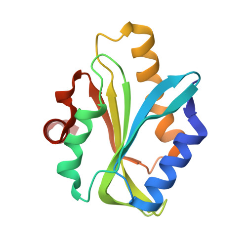 | |
UniProt | |||||
Find proteins for N9V330 (Entamoeba histolytica HM-1:IMSS-A) Explore N9V330 Go to UniProtKB: N9V330 | |||||
Entity Groups | |||||
| Sequence Clusters | 30% Identity50% Identity70% Identity90% Identity95% Identity100% Identity | ||||
| UniProt Group | N9V330 | ||||
Sequence AnnotationsExpand | |||||
| |||||
| Length ( Å ) | Angle ( ˚ ) |
|---|---|
| a = 34.71 | α = 90 |
| b = 86.544 | β = 90 |
| c = 88.06 | γ = 90 |
| Software Name | Purpose |
|---|---|
| REFMAC | refinement |
| HKL-2000 | data reduction |
| SCALEPACK | data scaling |
| PHASER | phasing |
RCSB PDB (citation) is hosted by
RCSB PDB is a member of the

