6J55

Crystal structure of an iron superoxide dismutate (FeSOD) from a pathogenic Acanthamoeba castellanii


Experimental Data Snapshot

  • Method: X-RAY DIFFRACTION
  • Resolution: 2.33 Å
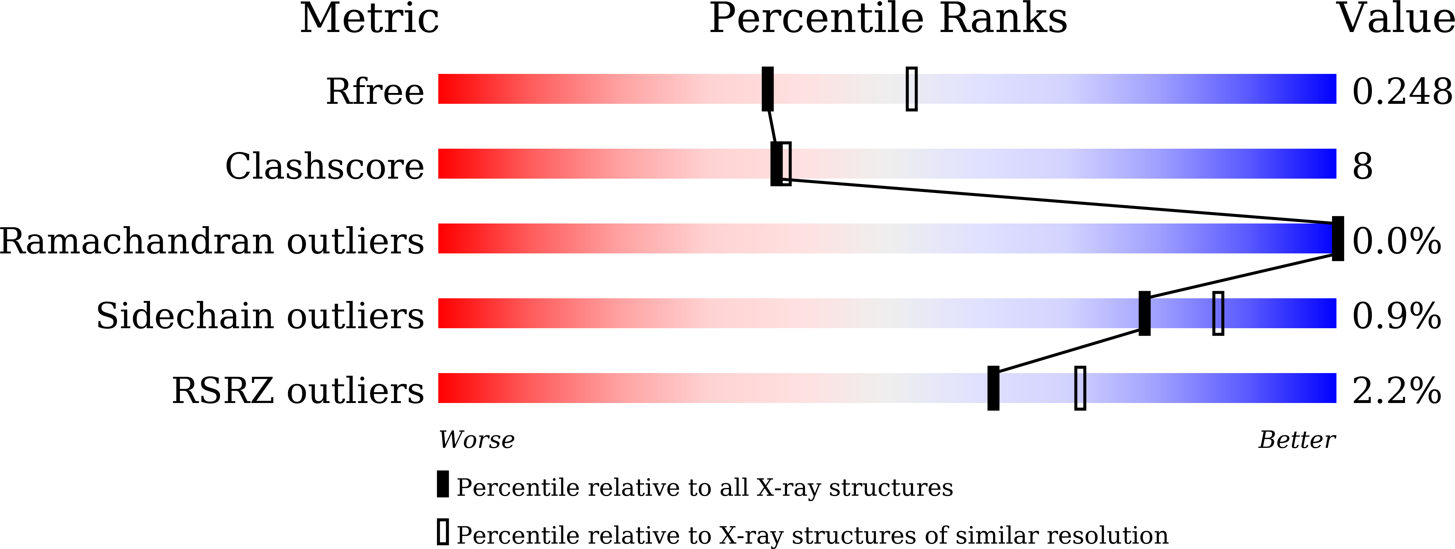
  • R-Value Free: 0.248 
  • R-Value Work: 0.200 
  • R-Value Observed: 0.202 

wwPDB Validation   3D Report Full Report


This is version 1.3 of the entry. See complete history


Literature

Crystal structure of an iron superoxide dismutase from the pathogenic amoeba Acanthamoeba castellanii.

Dao, O.Asaithambi, K.Na, B.K.Lee, K.H.

(2019) Acta Crystallogr F Struct Biol Commun 75: 480-488

  • DOI: https://doi.org/10.1107/S2053230X19008112
  • Primary Citation of Related Structures:  
    6J55

  • PubMed Abstract: 

    The iron superoxide dismutase found in the pathogenic amoeba Acanthamoeba castellanii (AcFeSOD) may play essential roles in the survival of the parasite, not only by protecting it from endogenous oxidative stress but also by detoxifying oxidative killing of the parasite by host immune effector cells. The AcFeSOD protein was expressed in a stable form using an Escherichia coli expression system and was crystallized by the microbatch and hanging-drop vapour-diffusion methods. The structure was determined to 2.33 Å resolution from a single AcFeSOD crystal. The crystal belonged to the hexagonal space group P6 1 and contained 12 molecules forming three tetramers in the asymmetric unit, with an iron ion bound in each molecule. Structural comparisons and sequence alignment of AcFeSOD with other FeSODs showed a well conserved overall fold and conserved active-site residues with subtle differences.


  • Organizational Affiliation

    Department of Convergence Medical Science, Gyeongsang National University College of Medicine, Jinju, Gyeongsangnam, Republic of Korea.


Macromolecules
Find similar proteins by:  (by identity cutoff)  |  3D Structure
Entity ID: 1
MoleculeChains Sequence LengthOrganismDetailsImage
Superoxide dismutase
A, B, C, D, E
A, B, C, D, E, F, G, H, I, J, K, L
205Acanthamoeba castellaniiMutation(s): 0 
EC: 1.15.1.1
UniProt
Find proteins for Q5IZD9 (Acanthamoeba castellanii)
Explore Q5IZD9 
Go to UniProtKB:  Q5IZD9
Entity Groups  
Sequence Clusters30% Identity50% Identity70% Identity90% Identity95% Identity100% Identity
UniProt GroupQ5IZD9
Sequence Annotations
Expand
  • Reference Sequence
Small Molecules
Ligands 1 Unique
IDChains Name / Formula / InChI Key2D Diagram3D Interactions
FE2
Query on FE2

Download Ideal Coordinates CCD File 
M [auth A]
N [auth B]
O [auth C]
P [auth D]
Q [auth E]
M [auth A],
N [auth B],
O [auth C],
P [auth D],
Q [auth E],
R [auth F],
S [auth G],
T [auth H],
U [auth I],
V [auth J],
W [auth K],
X [auth L]
FE (II) ION
Fe
CWYNVVGOOAEACU-UHFFFAOYSA-N
Experimental Data & Validation

Experimental Data

  • Method: X-RAY DIFFRACTION
  • Resolution: 2.33 Å
  • R-Value Free: 0.248 
  • R-Value Work: 0.200 
  • R-Value Observed: 0.202 
  • Space Group: P 61
Unit Cell:
Length ( Å )Angle ( ˚ )
a = 127.486α = 90
b = 127.486β = 90
c = 327.064γ = 120
Software Package:
Software NamePurpose
PHENIXrefinement
HKL-2000data reduction
HKL-2000data scaling
MOLREPphasing
PDB_EXTRACTdata extraction

Structure Validation

View Full Validation Report


Entry History & Funding Information

Deposition Data


Funding OrganizationLocationGrant Number
National Research Foundation (Korea)Korea, Republic OfNRF-2015R1D1A1A01060694
National Research Foundation (Korea)Korea, Republic OfNRF-2016M3D3A1A01913246

Revision History  (Full details and data files)

  • Version 1.0: 2019-07-17
    Type: Initial release
  • Version 1.1: 2019-11-20
    Changes: Database references
  • Version 1.2: 2023-11-22
    Changes: Data collection, Database references, Derived calculations, Refinement description
  • Version 1.3: 2024-03-20
    Changes: Source and taxonomy