6H3A

Crystal structure of the KAP1 RBCC domain in complex with the SMARCAD1 CUE1 domain.


Experimental Data Snapshot

  • Method: X-RAY DIFFRACTION
  • Resolution: 5.50 Å
  • R-Value Free: 0.303 
  • R-Value Work: 0.279 
  • R-Value Observed: 0.281 

wwPDB Validation   3D Report Full Report


This is version 1.1 of the entry. See complete history


Literature

A Ubiquitin-Binding Domain that Binds a Structural Fold Distinct from that of Ubiquitin.

Lim, M.Newman, J.A.Williams, H.L.Masino, L.Aitkenhead, H.Gravard, A.E.Gileadi, O.Svejstrup, J.Q.

(2019) Structure 27: 1316-1325.e6

  • DOI: https://doi.org/10.1016/j.str.2019.05.003
  • Primary Citation of Related Structures:  
    6H3A, 6QU1

  • PubMed Abstract: 

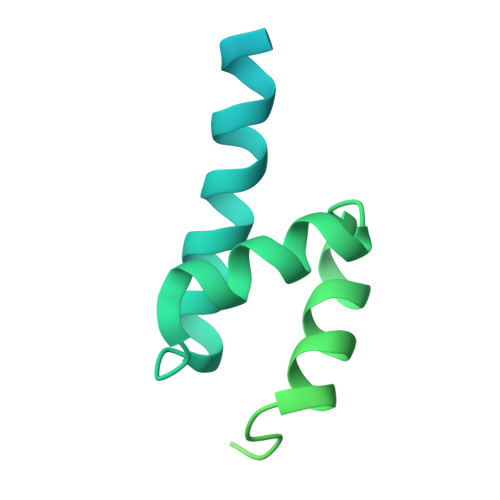
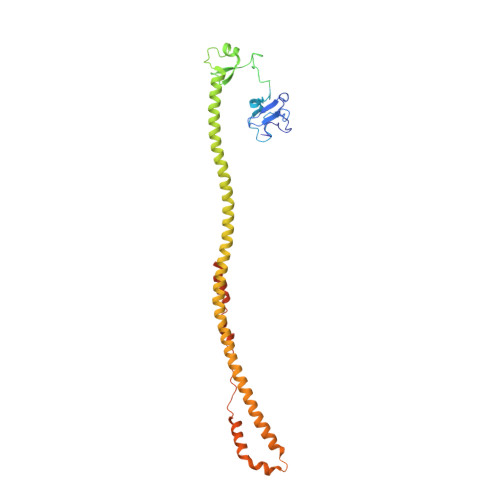
    Ubiquitylation, the posttranslational linkage of ubiquitin moieties to lysines in target proteins, helps regulate a myriad of biological processes. Ubiquitin, and sometimes ubiquitin-homology domains, are recognized by ubiquitin-binding domains, including CUE domains. CUE domains are thus generally thought to function by mediating interactions with ubiquitylated proteins. The chromatin remodeler, SMARCAD1, interacts with KAP1, a transcriptional corepressor. The SMARCAD1-KAP1 interaction is direct and involves the first SMARCAD1 CUE domain (CUE1) and the RBCC domain of KAP1. Here, we present a structural model of the KAP1 RBCC-SMARCAD1 CUE1 complex based on X-ray crystallography. Remarkably, CUE1, a canonical CUE domain, recognizes a cluster of exposed hydrophobic and surrounding charged/amphipathic residues on KAP1, which are presented in the context of a coiled-coil domain, not in a structure resembling ubiquitin. Together, these data suggest that CUE domains may have a wider function than simply recognizing ubiquitin and the ubiquitin-fold.


  • Organizational Affiliation

    Mechanisms of Transcription Laboratory, The Francis Crick Institute, 1 Midland Road, London NW1 1AT, UK.


Macromolecules
Find similar proteins by:  (by identity cutoff)  |  3D Structure
Entity ID: 1
MoleculeChains Sequence LengthOrganismDetailsImage
SWI/SNF-related matrix-associated actin-dependent regulator of chromatin subfamily A containing DEAD/H box 1A [auth D],
C [auth B]
253Homo sapiensMutation(s): 0 
Gene Names: SMARCAD1KIAA1122
EC: 3.6.4.12
UniProt & NIH Common Fund Data Resources
Find proteins for Q9H4L7 (Homo sapiens)
Explore Q9H4L7 
Go to UniProtKB:  Q9H4L7
PHAROS:  Q9H4L7
GTEx:  ENSG00000163104 
Entity Groups  
Sequence Clusters30% Identity50% Identity70% Identity90% Identity95% Identity100% Identity
UniProt GroupQ9H4L7
Sequence Annotations
Expand
  • Reference Sequence
Find similar proteins by:  (by identity cutoff)  |  3D Structure
Entity ID: 2
MoleculeChains Sequence LengthOrganismDetailsImage
Transcription intermediary factor 1-betaB [auth A],
D [auth F]
382Homo sapiensMutation(s): 0 
Gene Names: TRIM28KAP1RNF96TIF1B
EC: 2.3.2.27
UniProt & NIH Common Fund Data Resources
Find proteins for Q13263 (Homo sapiens)
Explore Q13263 
Go to UniProtKB:  Q13263
PHAROS:  Q13263
GTEx:  ENSG00000130726 
Entity Groups  
Sequence Clusters30% Identity50% Identity70% Identity90% Identity95% Identity100% Identity
UniProt GroupQ13263
Sequence Annotations
Expand
  • Reference Sequence
Experimental Data & Validation

Experimental Data

  • Method: X-RAY DIFFRACTION
  • Resolution: 5.50 Å
  • R-Value Free: 0.303 
  • R-Value Work: 0.279 
  • R-Value Observed: 0.281 
  • Space Group: I 2 3
Unit Cell:
Length ( Å )Angle ( ˚ )
a = 299.914α = 90
b = 299.914β = 90
c = 299.914γ = 90
Software Package:
Software NamePurpose
PHENIXrefinement
DIALSdata reduction
Aimlessdata scaling
AutoSolphasing

Structure Validation

View Full Validation Report


Entry History 

Deposition Data

Revision History  (Full details and data files)

  • Version 1.0: 2019-06-26
    Type: Initial release
  • Version 1.1: 2019-08-14
    Changes: Data collection, Database references