6F6J

Crystal structure of the Fe(II)/alpha-ketoglutarate dependent dioxygenase KDO1 with Fe(II)/succinate/(3S)-3-hydroxy-L-lysine


Experimental Data Snapshot

  • Method: X-RAY DIFFRACTION
  • Resolution: 2.00 Å
  • R-Value Free: 0.225 
  • R-Value Work: 0.194 
  • R-Value Observed: 0.196 

wwPDB Validation   3D Report Full Report


Ligand Structure Quality Assessment 


This is version 2.0 of the entry. See complete history


Literature

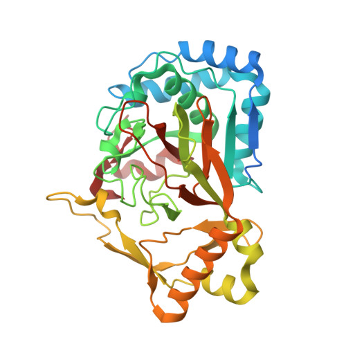
Structural Studies based on two Lysine Dioxygenases with Distinct Regioselectivity Brings Insights Into Enzyme Specificity within the Clavaminate Synthase-Like Family.

Bastard, K.Isabet, T.Stura, E.A.Legrand, P.Zaparucha, A.

(2018) Sci Rep 8: 16587-16587

  • DOI: https://doi.org/10.1038/s41598-018-34795-9
  • Primary Citation of Related Structures:  
    6EUO, 6EUR, 6EXF, 6EXH, 6F2A, 6F2B, 6F2E, 6F6J, 6F9P

  • PubMed Abstract: 

    Iron(II)/α-ketoacid-dependent oxygenases (αKAOs) are enzymes that catalyze the oxidation of unactivated C-H bonds, mainly through hydroxylation. Among these, those that are active towards amino-acids and their derivatives are grouped in the Clavaminate Synthase Like (CSL) family. CSL enzymes exhibit high regio- and stereoselectivities with strict substrate specificity. This study reports the structural elucidation of two new regiodivergent members, KDO1 and KDO5, active towards lysine, and the structural and computational analysis of the whole family through modelling and classification of active sites. The structures of KDO1 and KDO5 in complex with their ligands show that one exact position in the active site controls the regioselectivity of the reaction. Our results suggest that the substrate specificity and high stereoselectivity typical of this family is linked to a lid that closes up in order to form a sub-pocket around the side chain of the substrate. This dynamic lid is found throughout the family with varying sequence and length and is associated with a conserved stable dimeric interface. Results from this study could be a starting-point for exploring the functional diversity of the CSL family and direct in vitro screening in the search for new enzymatic activities.


  • Organizational Affiliation

    Génomique Métabolique, Genoscope, Institut François Jacob, CEA, CNRS, Univ Evry, Université Paris-Saclay, 91057, Evry, France.


Macromolecules
Find similar proteins by:  (by identity cutoff)  |  3D Structure
Entity ID: 1
MoleculeChains Sequence LengthOrganismDetailsImage
L-lysine 3-hydroxylase
A, B, C, D
358Catenulispora acidiphila DSM 44928Mutation(s): 0 
Gene Names: Caci_0231
EC: 1.14.11
UniProt
Find proteins for C7QJ42 (Catenulispora acidiphila (strain DSM 44928 / JCM 14897 / NBRC 102108 / NRRL B-24433 / ID139908))
Explore C7QJ42 
Go to UniProtKB:  C7QJ42
Entity Groups  
Sequence Clusters30% Identity50% Identity70% Identity90% Identity95% Identity100% Identity
UniProt GroupC7QJ42
Sequence Annotations
Expand
  • Reference Sequence
Small Molecules
Ligands 4 Unique
IDChains Name / Formula / InChI Key2D Diagram3D Interactions
CUW
Query on CUW

Download Ideal Coordinates CCD File 
G [auth A],
K [auth B],
N [auth C],
Q [auth D]
(2~{S},3~{R})-2,6-bis(azanyl)-3-oxidanyl-hexanoic acid
C6 H14 N2 O3
YSVMULOOWPBERR-UHNVWZDZSA-N
SIN
Query on SIN

Download Ideal Coordinates CCD File 
F [auth A],
J [auth B],
M [auth C],
P [auth D]
SUCCINIC ACID
C4 H6 O4
KDYFGRWQOYBRFD-UHFFFAOYSA-N
ACT
Query on ACT

Download Ideal Coordinates CCD File 
H [auth A],
R [auth D]
ACETATE ION
C2 H3 O2
QTBSBXVTEAMEQO-UHFFFAOYSA-M
FE
Query on FE

Download Ideal Coordinates CCD File 
E [auth A],
I [auth B],
L [auth C],
O [auth D]
FE (III) ION
Fe
VTLYFUHAOXGGBS-UHFFFAOYSA-N
Experimental Data & Validation

Experimental Data

  • Method: X-RAY DIFFRACTION
  • Resolution: 2.00 Å
  • R-Value Free: 0.225 
  • R-Value Work: 0.194 
  • R-Value Observed: 0.196 
  • Space Group: P 1
Unit Cell:
Length ( Å )Angle ( ˚ )
a = 56.95α = 107.55
b = 67.39β = 102.87
c = 110.65γ = 93.58
Software Package:
Software NamePurpose
BUSTERrefinement
XDSdata reduction
Cootmodel building
MOLREPphasing
XSCALEdata scaling

Structure Validation

View Full Validation Report


Ligand Structure Quality Assessment 


Entry History 

Deposition Data

Revision History  (Full details and data files)

  • Version 1.0: 2018-11-14
    Type: Initial release
  • Version 1.1: 2018-11-21
    Changes: Data collection, Database references
  • Version 2.0: 2023-11-15
    Changes: Atomic model, Data collection, Database references, Derived calculations