6ELC

Crystal Structure of O-linked Glycosylated VSG3

  • Classification: IMMUNE SYSTEM
  • Organism(s): Trypanosoma brucei brucei
  • Mutation(s): No 
  • Membrane Protein: Yes  mpstruc

  • Deposited: 2017-09-28 Released: 2018-07-11 
  • Deposition Author(s): Stebbins, C.E.
  • Funding Organization(s): National Institutes of Health/National Institute of General Medical Sciences (NIH/NIGMS), National Institutes of Health/National Institute Of Allergy and Infectious Diseases (NIH/NIAID), Wellcome Trust, National Institutes of Health

Experimental Data Snapshot

  • Method: X-RAY DIFFRACTION
  • Resolution: 1.41 Å
  • R-Value Free: 0.169 
  • R-Value Work: 0.130 
  • R-Value Observed: 0.132 

wwPDB Validation   3D Report Full Report


Ligand Structure Quality Assessment 


This is version 2.1 of the entry. See complete history

Re-refinement Note

A newer entry is available that reflects an alternative modeling of the original data: 7P59


Literature

African trypanosomes evade immune clearance by O-glycosylation of the VSG surface coat.

Pinger, J.Nesic, D.Ali, L.Aresta-Branco, F.Lilic, M.Chowdhury, S.Kim, H.S.Verdi, J.Raper, J.Ferguson, M.A.J.Papavasiliou, F.N.Stebbins, C.E.

(2018) Nat Microbiol 3: 932-938

  • DOI: https://doi.org/10.1038/s41564-018-0187-6
  • Primary Citation of Related Structures:  
    6ELC

  • PubMed Abstract: 

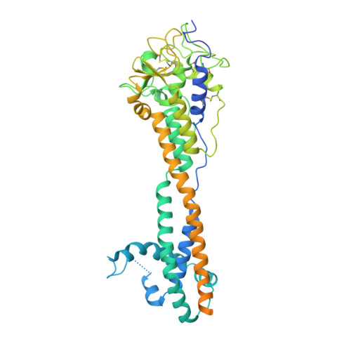
    The African trypanosome Trypanosoma brucei spp. is a paradigm for antigenic variation, the orchestrated alteration of cell surface molecules to evade host immunity. The parasite elicits robust antibody-mediated immune responses to its variant surface glycoprotein (VSG) coat, but evades immune clearance by repeatedly accessing a large genetic VSG repertoire and 'switching' to antigenically distinct VSGs. This persistent immune evasion has been ascribed exclusively to amino-acid variance on the VSG surface presented by a conserved underlying protein architecture. We establish here that this model does not account for the scope of VSG structural and biochemical diversity. The 1.4-Å-resolution crystal structure of the variant VSG3 manifests divergence in the tertiary fold and oligomeric state. The structure also reveals an O-linked carbohydrate on the top surface of VSG3. Mass spectrometric analysis indicates that this O-glycosylation site is heterogeneously occupied in VSG3 by zero to three hexose residues and is also present in other VSGs. We demonstrate that this O-glycosylation increases parasite virulence by impairing the generation of protective immunity. These data alter the paradigm of antigenic variation by the African trypanosome, expanding VSG variability beyond amino-acid sequence to include surface post-translational modifications with immunomodulatory impact.


  • Organizational Affiliation

    The Rockefeller University, Laboratory of Lymphocyte Biology, New York, NY, USA.


Macromolecules
Find similar proteins by:  (by identity cutoff)  |  3D Structure
Entity ID: 1
MoleculeChains Sequence LengthOrganismDetailsImage
Variant surface glycoprotein509Trypanosoma brucei bruceiMutation(s): 0 
Membrane Entity: Yes 
UniProt
Find proteins for B3GVK1 (Trypanosoma brucei brucei)
Explore B3GVK1 
Go to UniProtKB:  B3GVK1
Entity Groups  
Sequence Clusters30% Identity50% Identity70% Identity90% Identity95% Identity100% Identity
UniProt GroupB3GVK1
Sequence Annotations
Expand
  • Reference Sequence
Oligosaccharides

Help

Entity ID: 2
MoleculeChains Length2D Diagram Glycosylation3D Interactions
alpha-D-mannopyranose-(1-6)-beta-D-mannopyranose-(1-4)-2-acetamido-2-deoxy-beta-D-glucopyranose-(1-4)-2-acetamido-2-deoxy-beta-D-glucopyranose
B
4N-Glycosylation
Glycosylation Resources
GlyTouCan:  G22573RC
GlyCosmos:  G22573RC
GlyGen:  G22573RC
Small Molecules
Ligands 1 Unique
IDChains Name / Formula / InChI Key2D Diagram3D Interactions
GLC
Query on GLC

Download Ideal Coordinates CCD File 
C [auth A]alpha-D-glucopyranose
C6 H12 O6
WQZGKKKJIJFFOK-DVKNGEFBSA-N
Experimental Data & Validation

Experimental Data

  • Method: X-RAY DIFFRACTION
  • Resolution: 1.41 Å
  • R-Value Free: 0.169 
  • R-Value Work: 0.130 
  • R-Value Observed: 0.132 
  • Space Group: I 21 3
Unit Cell:
Length ( Å )Angle ( ˚ )
a = 129.854α = 90
b = 129.854β = 90
c = 129.854γ = 90
Software Package:
Software NamePurpose
REFMACrefinement
XDSdata reduction
XDSdata scaling

Structure Validation

View Full Validation Report


Ligand Structure Quality Assessment 


Entry History & Funding Information

Deposition Data


Funding OrganizationLocationGrant Number
National Institutes of Health/National Institute of General Medical Sciences (NIH/NIGMS)United StatesGM103403
National Institutes of Health/National Institute Of Allergy and Infectious Diseases (NIH/NIAID)United StatesAI085973
Wellcome TrustUnited Kingdom101842
Wellcome TrustUnited Kingdom097045
National Institutes of HealthUnited States--

Revision History  (Full details and data files)

  • Version 1.0: 2018-07-11
    Type: Initial release
  • Version 1.1: 2018-07-18
    Changes: Data collection, Database references
  • Version 1.2: 2018-08-29
    Changes: Data collection, Database references
  • Version 2.0: 2020-07-29
    Type: Remediation
    Reason: Carbohydrate remediation
    Changes: Atomic model, Data collection, Derived calculations, Structure summary
  • Version 2.1: 2022-03-30
    Changes: Author supporting evidence, Database references, Derived calculations, Structure summary