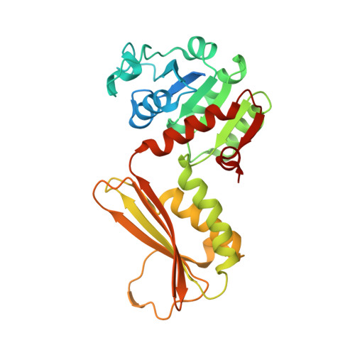
Plant DHDPR forms a dimer with unique secondary structure features that preclude higher-order assembly.
Watkin, S.A.J., Keown, J.R., Richards, E., Goldstone, D.C., Devenish, S.R.A., Grant Pearce, F.(2018) Biochem J 475: 137-150
- PubMed: 29187521
- DOI: https://doi.org/10.1042/BCJ20170709
- Primary Citation of Related Structures:
5U5I, 5U5N, 5UA0, 5UGJ - PubMed Abstract:
Dihydrodipicolinate reductase (DHDPR) catalyses the second reaction in the diaminopimelate pathway of lysine biosynthesis in bacteria and plants. In contrast with the tetrameric bacterial DHDPR enzymes, we show that DHDPR from Vitis vinifera (grape) and Selaginella moellendorffii are dimeric in solution. In the present study, we have also determined the crystal structures of DHDPR enzymes from the plants Arabidopsis thaliana and S. moellendorffii , which are the first dimeric DHDPR structures. The analysis of these models demonstrates that the dimer forms through the intra-strand interface, and that unique secondary features in the plant enzymes block tetramer assembly. In addition, we have also solved the structure of tetrameric DHDPR from the pathogenic bacteria Neisseria meningitidis Measuring the activity of plant DHDPR enzymes showed that they are much more prone to substrate inhibition than the bacterial enzymes, which appears to be a consequence of increased flexibility of the substrate-binding loop and higher affinity for the nucleotide substrate. This higher propensity to substrate inhibition may have consequences for ongoing efforts to increase lysine biosynthesis in plants.
Organizational Affiliation:
Biomolecular Interaction Centre and School of Biological Sciences, University of Canterbury, Christchurch, New Zealand.


