


Crystal structure of 2-methylcitrate dehydratase (MmgE) from Bacillus subtilis.
Baker, G.E., Race, P.R.To be published.
Experimental Data Snapshot
wwPDB Validation 3D Report Full Report

Entity ID: 1 | |||||
|---|---|---|---|---|---|
| Molecule | Chains | Sequence Length | Organism | Details | Image |
| 2-methylcitrate dehydratase | 472 | Bacillus subtilis subsp. subtilis str. 168 | Mutation(s): 0 Gene Names: prpD, mmgE, yqiP, BSU24130 EC: 4.2.1.79 (PDB Primary Data), 4.2.1.3 (PDB Primary Data) | 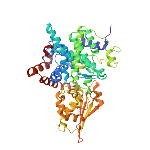 | |
UniProt | |||||
Find proteins for P45859 (Bacillus subtilis (strain 168)) Explore P45859 Go to UniProtKB: P45859 | |||||
Entity Groups | |||||
| Sequence Clusters | 30% Identity50% Identity70% Identity90% Identity95% Identity100% Identity | ||||
| UniProt Group | P45859 | ||||
Sequence AnnotationsExpand | |||||
| |||||
| Ligands 1 Unique | |||||
|---|---|---|---|---|---|
| ID | Chains | Name / Formula / InChI Key | 2D Diagram | 3D Interactions | |
| TLA Query on TLA | G [auth A] H [auth B] I [auth C] J [auth D] K [auth E] | L(+)-TARTARIC ACID C4 H6 O6 FEWJPZIEWOKRBE-JCYAYHJZSA-N |  | ||
| Length ( Å ) | Angle ( ˚ ) |
|---|---|
| a = 90.831 | α = 90 |
| b = 194.409 | β = 92.74 |
| c = 90.869 | γ = 90 |
| Software Name | Purpose |
|---|---|
| REFMAC | refinement |
| XDS | data reduction |
| Aimless | data scaling |
| PHASER | phasing |
RCSB PDB (citation) is hosted by
RCSB PDB is a member of the

