


Structural basis of p63/p73 hetero-tetramerization
Gebel, J., Luh, L., Coutandin, D., Lohr, F., Schafer, B., Sumyk, M., Buchner, L., Krojer, T., Salah, E., Mathea, S., Guntert, P., Knapp, S., Dotsch, V.To be published.
Experimental Data Snapshot
wwPDB Validation 3D Report Full Report

Entity ID: 1 | |||||
|---|---|---|---|---|---|
| Molecule | Chains | Sequence Length | Organism | Details | Image |
| Tumor protein p73 | 50 | Homo sapiens | Mutation(s): 4 Gene Names: TP73, P73 | 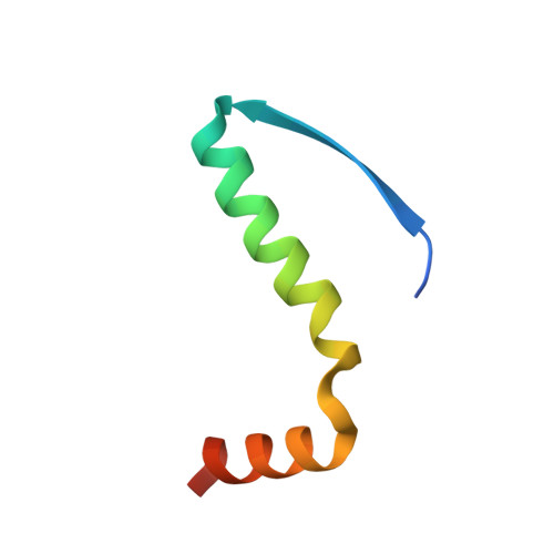 | |
UniProt & NIH Common Fund Data Resources | |||||
Find proteins for O15350 (Homo sapiens) Explore O15350 Go to UniProtKB: O15350 | |||||
PHAROS: O15350 GTEx: ENSG00000078900 | |||||
Entity Groups | |||||
| Sequence Clusters | 30% Identity50% Identity70% Identity90% Identity95% Identity100% Identity | ||||
| UniProt Group | O15350 | ||||
Sequence AnnotationsExpand | |||||
| |||||
| Ligands 1 Unique | |||||
|---|---|---|---|---|---|
| ID | Chains | Name / Formula / InChI Key | 2D Diagram | 3D Interactions | |
| MG Query on MG | I [auth F] | MAGNESIUM ION Mg JLVVSXFLKOJNIY-UHFFFAOYSA-N |  | ||
| Length ( Å ) | Angle ( ˚ ) |
|---|---|
| a = 29.05 | α = 89.6 |
| b = 48.4 | β = 83.2 |
| c = 75.73 | γ = 74.27 |
| Software Name | Purpose |
|---|---|
| XSCALE | data scaling |
| PHASER | phasing |
| PHENIX | refinement |
| PDB_EXTRACT | data extraction |
| XDS | data reduction |
RCSB PDB (citation) is hosted by
RCSB PDB is a member of the

