5BV3

Yeast Scavenger Decapping Enzyme in complex with m7GDP


Experimental Data Snapshot

  • Method: X-RAY DIFFRACTION
  • Resolution: 2.25 Å
  • R-Value Free: 0.248 
  • R-Value Work: 0.222 
  • R-Value Observed: 0.223 

wwPDB Validation   3D Report Full Report


Ligand Structure Quality Assessment 


This is version 1.3 of the entry. See complete history


Literature

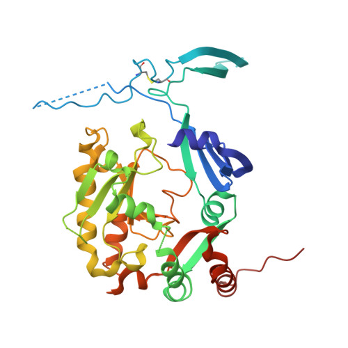
An excess of catalytically required motions inhibits the scavenger decapping enzyme.

Neu, A.Neu, U.Fuchs, A.L.Schlager, B.Sprangers, R.

(2015) Nat Chem Biol 11: 697-704

  • DOI: https://doi.org/10.1038/nchembio.1866
  • Primary Citation of Related Structures:  
    5BV3

  • PubMed Abstract: 

    The scavenger decapping enzyme hydrolyzes the protective 5' cap structure on short mRNA fragments that are generated from the exosomal degradation of mRNAs. From static crystal structures and NMR data, it is apparent that the dimeric enzyme has to undergo large structural changes to bind its substrate in a catalytically competent conformation. Here we studied the yeast enzyme and showed that the associated opening and closing motions can be orders of magnitude faster than the catalytic turnover rate. This excess of motion is induced by the binding of a second ligand to the enzyme, which occurs at high substrate concentrations. We designed a mutant that disrupted the allosteric pathway that links the second binding event to the dynamics and showed that this mutant enzyme is hyperactive. Our data reveal a unique mechanism of substrate inhibition in which motions that are required for catalytic activity also inhibit efficient turnover when they are present in excess.


  • Organizational Affiliation

    Max Planck Institute for Developmental Biology, Tübingen, Germany.


Macromolecules
Find similar proteins by:  (by identity cutoff)  |  3D Structure
Entity ID: 1
MoleculeChains Sequence LengthOrganismDetailsImage
m7GpppX diphosphatase
A, B, C, D
345Saccharomyces cerevisiae S288CMutation(s): 1 
Gene Names: DCS1YLR270W
EC: 3.6.1.59
UniProt
Find proteins for Q06151 (Saccharomyces cerevisiae (strain ATCC 204508 / S288c))
Explore Q06151 
Go to UniProtKB:  Q06151
Entity Groups  
Sequence Clusters30% Identity50% Identity70% Identity90% Identity95% Identity100% Identity
UniProt GroupQ06151
Sequence Annotations
Expand
  • Reference Sequence
Small Molecules
Ligands 3 Unique
IDChains Name / Formula / InChI Key2D Diagram3D Interactions
M7G
Query on M7G

Download Ideal Coordinates CCD File 
E [auth A],
K [auth C]
7N-METHYL-8-HYDROGUANOSINE-5'-DIPHOSPHATE
C11 H18 N5 O11 P2
SBASPRRECYVBRF-KQYNXXCUSA-O
SO4
Query on SO4

Download Ideal Coordinates CCD File 
F [auth A]
G [auth A]
I [auth B]
J [auth B]
L [auth C]
F [auth A],
G [auth A],
I [auth B],
J [auth B],
L [auth C],
N [auth D],
O [auth D]
SULFATE ION
O4 S
QAOWNCQODCNURD-UHFFFAOYSA-L
CL
Query on CL

Download Ideal Coordinates CCD File 
H [auth A],
M [auth C],
P [auth D],
Q [auth D]
CHLORIDE ION
Cl
VEXZGXHMUGYJMC-UHFFFAOYSA-M
Binding Affinity Annotations 
IDSourceBinding Affinity
M7G Binding MOAD:  5BV3 Kd: 68 (nM) from 1 assay(s)
Experimental Data & Validation

Experimental Data

  • Method: X-RAY DIFFRACTION
  • Resolution: 2.25 Å
  • R-Value Free: 0.248 
  • R-Value Work: 0.222 
  • R-Value Observed: 0.223 
  • Space Group: P 21 21 21
Unit Cell:
Length ( Å )Angle ( ˚ )
a = 87.99α = 90
b = 104.52β = 90
c = 189.96γ = 90
Software Package:
Software NamePurpose
REFMACrefinement
XSCALEdata scaling
PHASERphasing
PDB_EXTRACTdata extraction
XDSdata reduction

Structure Validation

View Full Validation Report


Ligand Structure Quality Assessment 


Entry History & Funding Information

Deposition Data


Funding OrganizationLocationGrant Number
Germany--

Revision History  (Full details and data files)

  • Version 1.0: 2015-08-12
    Type: Initial release
  • Version 1.1: 2015-08-19
    Changes: Database references
  • Version 1.2: 2015-08-26
    Changes: Database references
  • Version 1.3: 2024-01-10
    Changes: Data collection, Database references, Refinement description