5NMD

868 TCR Specific for HLA A02 presenting HIV Epitope SLYNTVATL


Experimental Data Snapshot

  • Method: X-RAY DIFFRACTION
  • Resolution: 2.07 Å
  • R-Value Free: 0.223 
  • R-Value Work: 0.189 
  • R-Value Observed: 0.190 

wwPDB Validation   3D Report Full Report


This is version 1.3 of the entry. See complete history


Literature

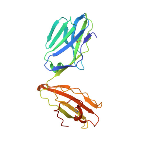
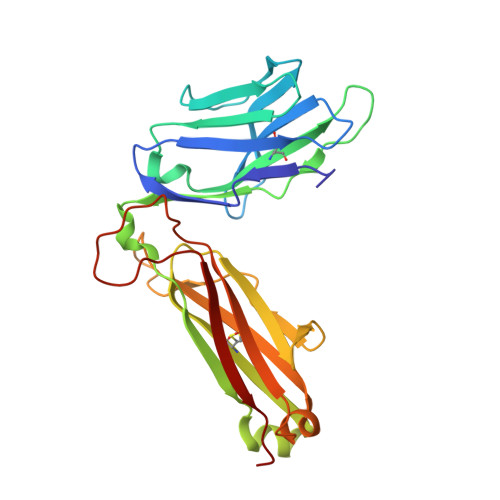
Dual Molecular Mechanisms Govern Escape at Immunodominant HLA A2-Restricted HIV Epitope.

Cole, D.K.Fuller, A.Dolton, G.Zervoudi, E.Legut, M.Miles, K.Blanchfield, L.Madura, F.Holland, C.J.Bulek, A.M.Bridgeman, J.S.Miles, J.J.Schauenburg, A.J.A.Beck, K.Evavold, B.D.Rizkallah, P.J.Sewell, A.K.

(2017) Front Immunol 8: 1503-1503

  • DOI: https://doi.org/10.3389/fimmu.2017.01503
  • Primary Citation of Related Structures:  
    5NMD, 5NME, 5NMF, 5NMG, 5NMH, 5NMK

  • PubMed Abstract: 

    Serial accumulation of mutations to fixation in the SLYNTVATL (SL9) immunodominant, HIV p17 Gag-derived, HLA A2-restricted cytotoxic T lymphocyte epitope produce the SLFNTIAVL triple mutant "ultimate" escape variant. These mutations in solvent-exposed residues are believed to interfere with TCR recognition, although confirmation has awaited structural verification. Here, we solved a TCR co-complex structure with SL9 and the triple escape mutant to determine the mechanism of immune escape in this eminent system. We show that, in contrast to prevailing hypotheses, the main TCR contact residue is 4N and the dominant mechanism of escape is not via lack of TCR engagement. Instead, mutation of solvent-exposed residues in the peptide destabilise the peptide-HLA and reduce peptide density at the cell surface. These results highlight the extraordinary lengths that HIV employs to evade detection by high-affinity TCRs with a broad peptide-binding footprint and necessitate re-evaluation of this exemplar model of HIV TCR escape.


  • Organizational Affiliation

    Cardiff University School of Medicine, University Hospital, Heath Park, Cardiff, United Kingdom.


Macromolecules
Find similar proteins by:  (by identity cutoff)  |  3D Structure
Entity ID: 1
MoleculeChains Sequence LengthOrganismDetailsImage
human T-cell Receptor alpha chain
A, C
200Homo sapiensMutation(s): 0 
Entity Groups  
Sequence Clusters30% Identity50% Identity70% Identity90% Identity95% Identity100% Identity
Sequence Annotations
Expand
  • Reference Sequence
Find similar proteins by:  (by identity cutoff)  |  3D Structure
Entity ID: 2
MoleculeChains Sequence LengthOrganismDetailsImage
Human T-cell Receptor, beta chain
B, D
242Homo sapiensMutation(s): 0 
Entity Groups  
Sequence Clusters30% Identity50% Identity70% Identity90% Identity95% Identity100% Identity
Sequence Annotations
Expand
  • Reference Sequence
Experimental Data & Validation

Experimental Data

  • Method: X-RAY DIFFRACTION
  • Resolution: 2.07 Å
  • R-Value Free: 0.223 
  • R-Value Work: 0.189 
  • R-Value Observed: 0.190 
  • Space Group: P 1 21 1
Unit Cell:
Length ( Å )Angle ( ˚ )
a = 87.47α = 90
b = 50.62β = 90.16
c = 114.31γ = 90
Software Package:
Software NamePurpose
REFMACrefinement
xia2data reduction
XDSdata reduction
Aimlessdata scaling
PHASERphasing

Structure Validation

View Full Validation Report


Entry History 

Deposition Data

Revision History  (Full details and data files)

  • Version 1.0: 2017-11-15
    Type: Initial release
  • Version 1.1: 2017-11-22
    Changes: Database references
  • Version 1.2: 2017-12-20
    Changes: Database references
  • Version 1.3: 2024-01-17
    Changes: Data collection, Database references, Refinement description