4XWW

Crystal structure of RNase J complexed with RNA


Experimental Data Snapshot

  • Method: X-RAY DIFFRACTION
  • Resolution: 1.70 Å
  • R-Value Free: 0.217 
  • R-Value Work: 0.195 
  • R-Value Observed: 0.196 

wwPDB Validation   3D Report Full Report


This is version 1.1 of the entry. See complete history


Literature

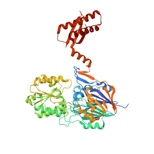
Structural insights into catalysis and dimerization enhanced exonuclease activity of RNase J

Zhao, Y.Lu, M.Zhang, H.Hu, J.Zhou, C.Xu, Q.Shah, A.M.U.H.Xu, H.Wang, L.Hua, Y.

(2015) Nucleic Acids Res 43: 5550-5559

  • DOI: https://doi.org/10.1093/nar/gkv444
  • Primary Citation of Related Structures:  
    4XWT, 4XWW

  • PubMed Abstract: 

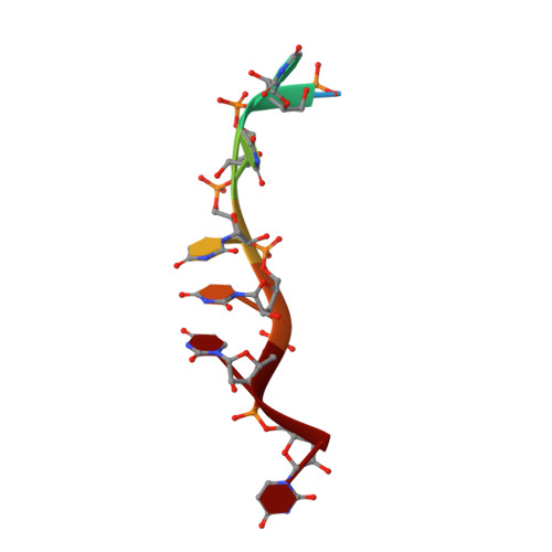
    RNase J is a conserved ribonuclease that belongs to the β-CASP family of nucleases. It possesses both endo- and exo-ribonuclease activities, which play a key role in pre-rRNA maturation and mRNA decay. Here we report high-resolution crystal structures of Deinococcus radiodurans RNase J complexed with RNA or uridine 5'-monophosphate in the presence of manganese ions. Biochemical and structural studies revealed that RNase J uses zinc ions for two-metal-ion catalysis. One residue conserved among RNase J orthologues (motif B) forms specific electrostatic interactions with the scissile phosphate of the RNA that is critical for the catalysis and product stabilization. The additional manganese ion, which is coordinated by conserved residues at the dimer interface, is critical for RNase J dimerization and exonuclease activity. The structures may also shed light on the mechanism of RNase J exo- and endonucleolytic activity switch.


  • Organizational Affiliation

    Key Laboratory of Chinese Ministry of Agriculture for Nuclear-Agricultural Sciences, Institute of Nuclear-Agricultural Sciences, Zhejiang University, China yezhao@zju.edu.cn.


Macromolecules

Find similar proteins by:  (by identity cutoff)  |  3D Structure
Entity ID: 1
MoleculeChains Sequence LengthOrganismDetailsImage
DR2417
A, B
559Deinococcus radioduransMutation(s): 0 
UniProt
Find proteins for H9CZL7 (Deinococcus radiodurans)
Explore H9CZL7 
Go to UniProtKB:  H9CZL7
Entity Groups  
Sequence Clusters30% Identity50% Identity70% Identity90% Identity95% Identity100% Identity
UniProt GroupH9CZL7
Sequence Annotations
Expand
  • Reference Sequence

Find similar nucleic acids by:  Sequence   |   3D Structure  

Entity ID: 2
MoleculeChains LengthOrganismImage
RNA (5'-D(UP*UP*UP*UP*UP*UP*U)-3')C [auth D],
D [auth E]
7synthetic construct
Sequence Annotations
Expand
  • Reference Sequence
Experimental Data & Validation

Experimental Data

  • Method: X-RAY DIFFRACTION
  • Resolution: 1.70 Å
  • R-Value Free: 0.217 
  • R-Value Work: 0.195 
  • R-Value Observed: 0.196 
  • Space Group: P 21 21 21
Unit Cell:
Length ( Å )Angle ( ˚ )
a = 67.12α = 90
b = 87.86β = 90
c = 249.4γ = 90
Software Package:
Software NamePurpose
PHENIXrefinement
XSCALEdata reduction
PDB_EXTRACTdata extraction
Cootmodel building
XDSphasing

Structure Validation

View Full Validation Report


Entry History & Funding Information

Deposition Data


Funding OrganizationLocationGrant Number
National Basic Research Program of ChinaChina2015CB910600

Revision History  (Full details and data files)

  • Version 1.0: 2015-12-16
    Type: Initial release
  • Version 1.1: 2024-03-20
    Changes: Data collection, Database references, Derived calculations