4XST

Structure of the endoglycosidase-H treated L1-CR domains of the human insulin receptor in complex with residues 697-719 of the human insulin receptor (A-isoform)


Experimental Data Snapshot

  • Method: X-RAY DIFFRACTION
  • Resolution: 3.00 Å
  • R-Value Free: 0.262 
  • R-Value Work: 0.217 
  • R-Value Observed: 0.219 

wwPDB Validation   3D Report Full Report


Ligand Structure Quality Assessment 


This is version 2.1 of the entry. See complete history


Literature

Structural Congruency of Ligand Binding to the Insulin and Insulin/Type 1 Insulin-like Growth Factor Hybrid Receptors.

Menting, J.G.Lawrence, C.F.Kong, G.K.Margetts, M.B.Ward, C.W.Lawrence, M.C.

(2015) Structure 23: 1271-1282

  • DOI: https://doi.org/10.1016/j.str.2015.04.016
  • Primary Citation of Related Structures:  
    4XSS, 4XST

  • PubMed Abstract: 

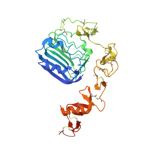
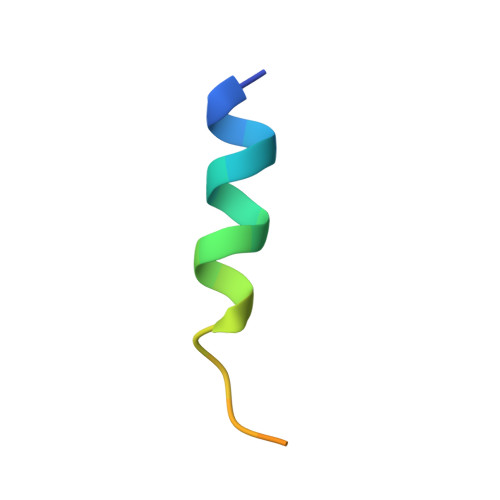
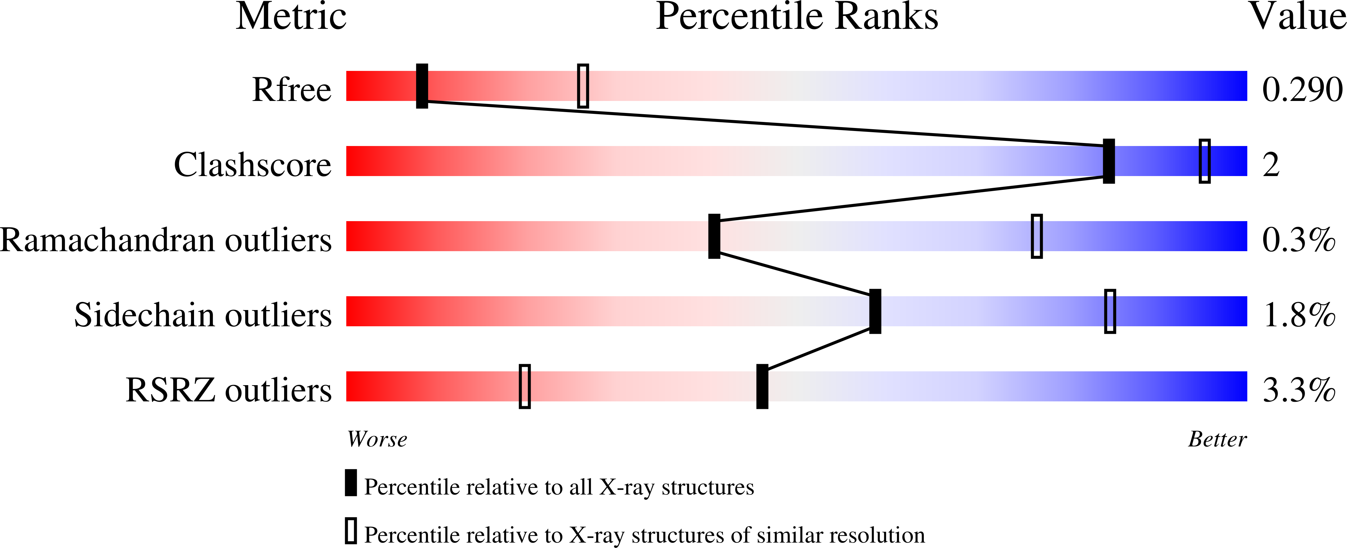
    The homodimeric insulin and type 1 insulin-like growth factor receptors (IR and IGF-1R) share a common architecture and each can bind all three ligands within the family: insulin and insulin-like growth factors I and II (IGF-I and IFG-II). The receptor monomers also assemble as heterodimers, the primary ligand-binding sites of which each comprise the first leucine-rich repeat domain (L1) of one receptor type and an α-chain C-terminal segment (αCT) of the second receptor type. We present here crystal structures of IGF-I bound to such a hybrid primary binding site and of a ligand-free version of an IR αCT peptide bound to an IR L1 plus cysteine-rich domain construct (IR310.T). These structures, refined at 3.0-Å resolution, prove congruent to respective existing structures of insulin-complexed IR310.T and the intact apo-IR ectodomain. As such, they provide key missing links in the emerging, but sparse, repertoire of structures defining the receptor family.


  • Organizational Affiliation

    Structural Biology Division, Walter and Eliza Hall Institute of Medical Research, Parkville, VIC 3052, Australia.


Macromolecules
Find similar proteins by:  (by identity cutoff)  |  3D Structure
Entity ID: 1
MoleculeChains Sequence LengthOrganismDetailsImage
Insulin receptorA [auth E]317Homo sapiensMutation(s): 0 
Gene Names: INSR
EC: 2.7.10.1
UniProt & NIH Common Fund Data Resources
Find proteins for P06213 (Homo sapiens)
Explore P06213 
Go to UniProtKB:  P06213
PHAROS:  P06213
GTEx:  ENSG00000171105 
Entity Groups  
Sequence Clusters30% Identity50% Identity70% Identity90% Identity95% Identity100% Identity
UniProt GroupP06213
Sequence Annotations
Expand
  • Reference Sequence

Find similar proteins by:  Sequence   |   3D Structure  

Entity ID: 2
MoleculeChains Sequence LengthOrganismDetailsImage
Insulin receptorB [auth F]23Rattus norvegicusMutation(s): 0 
EC: 2.7.10.1
UniProt
Find proteins for P15127 (Rattus norvegicus)
Explore P15127 
Go to UniProtKB:  P15127
Entity Groups  
Sequence Clusters30% Identity50% Identity70% Identity90% Identity95% Identity100% Identity
UniProt GroupP15127
Sequence Annotations
Expand
  • Reference Sequence
Oligosaccharides

Help

Entity ID: 3
MoleculeChains Length2D Diagram Glycosylation3D Interactions
alpha-L-fucopyranose-(1-6)-2-acetamido-2-deoxy-beta-D-glucopyranoseC [auth A]2N-Glycosylation
Glycosylation Resources
GlyTouCan:  G86851RC
GlyCosmos:  G86851RC
GlyGen:  G86851RC
Experimental Data & Validation

Experimental Data

  • Method: X-RAY DIFFRACTION
  • Resolution: 3.00 Å
  • R-Value Free: 0.262 
  • R-Value Work: 0.217 
  • R-Value Observed: 0.219 
  • Space Group: P 63 2 2
Unit Cell:
Length ( Å )Angle ( ˚ )
a = 158.718α = 90
b = 158.718β = 90
c = 85.912γ = 120
Software Package:
Software NamePurpose
BUSTERrefinement
Cootmodel building
PHASERphasing
XSCALEdata scaling
XDSdata reduction
Blu-Icedata collection

Structure Validation

View Full Validation Report


Ligand Structure Quality Assessment 


Entry History & Funding Information

Deposition Data


Funding OrganizationLocationGrant Number
National Health and Medical Research Council (NHMRC, Australia)AustraliaAPP1005896
National Health and Medical Research Council (NHMRC, Australia)AustraliaAPP1058233

Revision History  (Full details and data files)

  • Version 1.0: 2015-06-10
    Type: Initial release
  • Version 1.1: 2015-07-22
    Changes: Database references
  • Version 1.2: 2017-09-13
    Changes: Author supporting evidence, Data collection, Database references, Derived calculations, Source and taxonomy
  • Version 1.3: 2020-01-08
    Changes: Author supporting evidence, Data collection
  • Version 2.0: 2020-07-29
    Type: Remediation
    Reason: Carbohydrate remediation
    Changes: Atomic model, Data collection, Derived calculations, Structure summary
  • Version 2.1: 2023-09-27
    Changes: Data collection, Database references, Derived calculations, Refinement description, Structure summary