4IBN

Crystal structure of LC9-RNase H1, a type 1 RNase H with the type 2 active-site motif


Experimental Data Snapshot

  • Method: X-RAY DIFFRACTION
  • Resolution: 1.62 Å
  • R-Value Free: 0.215 
  • R-Value Work: 0.177 
  • R-Value Observed: 0.179 

wwPDB Validation   3D Report Full Report


This is version 1.3 of the entry. See complete history


Literature

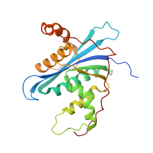
Crystal structure of metagenome-derived LC9-RNase H1 with atypical DEDN active site motif

Nguyen, T.-N.You, D.-J.Kanaya, E.Koga, Y.Kanaya, S.

(2013) FEBS Lett 587: 1418-1423

  • DOI: https://doi.org/10.1016/j.febslet.2013.03.020
  • Primary Citation of Related Structures:  
    4IBN

  • PubMed Abstract: 

    The crystal structure of metagenome-derived LC9-RNase H1 was determined. The structure-based mutational analyses indicated that the active site motif of LC9-RNase H1 is altered from DEDD to DEDN. In this motif, the location of the second glutamate residue is moved from αA-helix to β1-strand immediately next to the first aspartate residue, as in the active site of RNase H2. However, the structure and enzymatic properties of LC9-RNase H1 highly resemble those of RNase H1, instead of RNase H2. We propose that LC9-RNase H1 represents bacterial RNases H1 with an atypical DEDN active site motif, which are evolutionarily distinct from those with a typical DEDD active site motif.


  • Organizational Affiliation

    Department of Material and Life Science, Graduate School of Engineering, Osaka University, 2-1 Yamadaoka, Suita, Osaka 565-0871, Japan.


Macromolecules
Find similar proteins by:  (by identity cutoff)  |  3D Structure
Entity ID: 1
MoleculeChains Sequence LengthOrganismDetailsImage
Ribonuclease H220uncultured organismMutation(s): 0 
EC: 3.1.26.4
UniProt
Find proteins for E0X765 (uncultured organism)
Explore E0X765 
Go to UniProtKB:  E0X765
Entity Groups  
Sequence Clusters30% Identity50% Identity70% Identity90% Identity95% Identity100% Identity
UniProt GroupE0X765
Sequence Annotations
Expand
  • Reference Sequence
Experimental Data & Validation

Experimental Data

  • Method: X-RAY DIFFRACTION
  • Resolution: 1.62 Å
  • R-Value Free: 0.215 
  • R-Value Work: 0.177 
  • R-Value Observed: 0.179 
  • Space Group: P 1 21 1
Unit Cell:
Length ( Å )Angle ( ˚ )
a = 41.633α = 90
b = 39.913β = 102.64
c = 50.254γ = 90
Software Package:
Software NamePurpose
DENZOdata reduction
SCALEPACKdata scaling
MOLREPphasing
REFMACrefinement
PDB_EXTRACTdata extraction
HKL-2000data collection

Structure Validation

View Full Validation Report


Entry History 

Deposition Data

Revision History  (Full details and data files)

  • Version 1.0: 2013-03-20
    Type: Initial release
  • Version 1.1: 2013-07-10
    Changes: Database references
  • Version 1.2: 2017-11-15
    Changes: Refinement description
  • Version 1.3: 2023-11-08
    Changes: Data collection, Database references, Refinement description