4DNN

Crystal structure of the Quaking Qua1 homodimerization domain


Experimental Data Snapshot

  • Method: X-RAY DIFFRACTION
  • Resolution: 2.10 Å
  • R-Value Free: 0.252 
  • R-Value Work: 0.216 
  • R-Value Observed: 0.218 

wwPDB Validation   3D Report Full Report


This is version 1.1 of the entry. See complete history


Literature

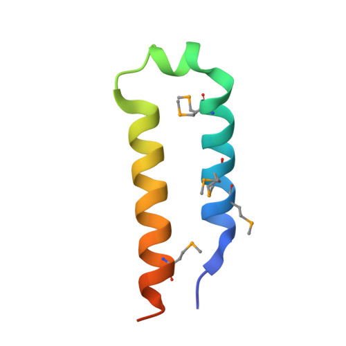
Structural Analysis of the Quaking Homodimerization Interface.

Beuck, C.Qu, S.Fagg, W.S.Ares, M.Williamson, J.R.

(2012) J Mol Biol 423: 766-781

  • DOI: https://doi.org/10.1016/j.jmb.2012.08.027
  • Primary Citation of Related Structures:  
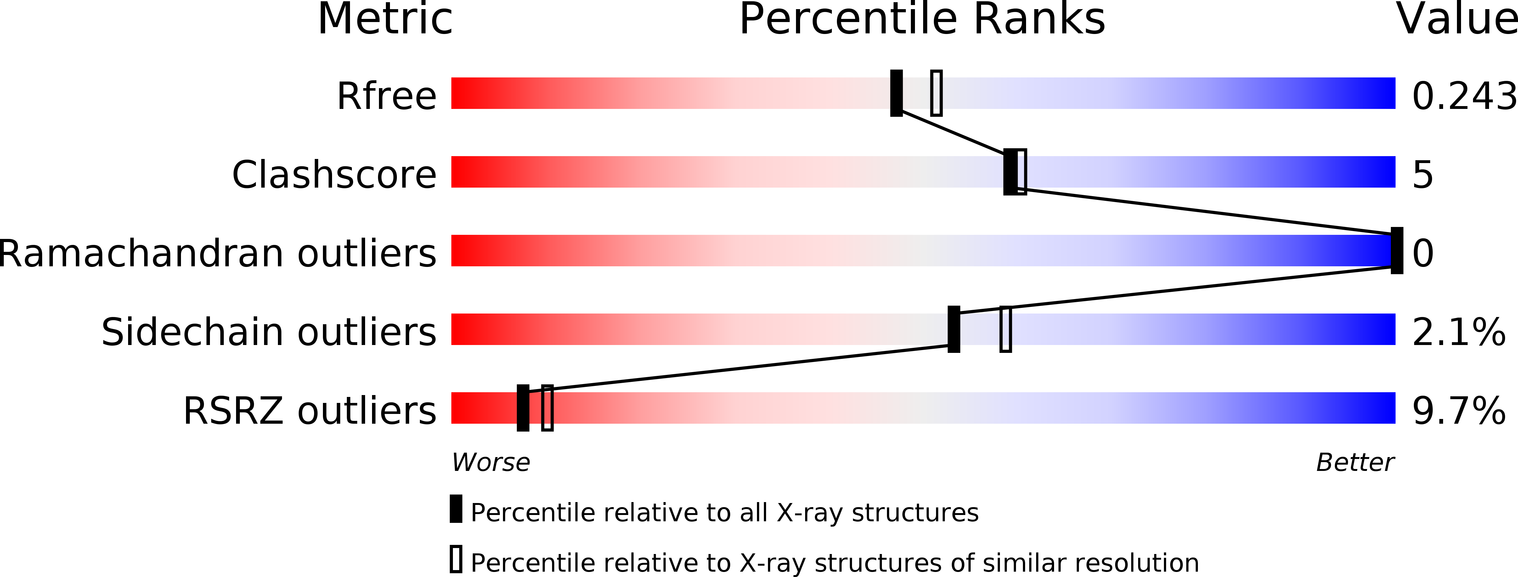
    4DNN

  • PubMed Abstract: 

    Quaking (QkI) is a prototypical member of the STAR (signal transducer and activator of RNA) protein family, which plays key roles in posttranscriptional gene regulation by controlling mRNA translation, stability and splicing. QkI-5 has been shown to regulate mRNA expression in the central nervous system, but little is known about its roles in other tissues. STAR proteins function as dimers and bind to bipartite RNA sequences; however, the structural and functional roles of homodimerization and heterodimerization are still unclear. Here, we present the crystal structure of the QkI dimerization domain, which adopts a similar stacked helix-turn-helix arrangement as its homologs GLD-1 (germ line development defective-1) and Sam68 (Src-associated protein during mitosis, 68kDa) but differs by an additional helix inserted in the dimer interface. Variability of the dimer interface residues likely ensures selective homodimerization by preventing association with non-cognate STAR family proteins in the cell. Mutations that inhibit dimerization also significantly impair RNA binding in vitro, alter QkI-5 protein levels and impair QkI function in a splicing assay in vivo. Together, our results indicate that a functional Qua1 homodimerization domain is required for QkI-5 function in mammalian cells.


  • Organizational Affiliation

    Department of Molecular Biology, Department of Chemistry and The Skaggs Institute for Chemical Biology, The Scripps Research Institute, La Jolla, CA 92037, USA.


Macromolecules
Find similar proteins by:  (by identity cutoff)  |  3D Structure
Entity ID: 1
MoleculeChains Sequence LengthOrganismDetailsImage
Protein quaking
A, B
56Mus musculusMutation(s): 1 
Gene Names: QkQk1Qka1QkiQuaking
UniProt
Find proteins for Q9QYS9 (Mus musculus)
Explore Q9QYS9 
Go to UniProtKB:  Q9QYS9
Entity Groups  
Sequence Clusters30% Identity50% Identity70% Identity90% Identity95% Identity100% Identity
UniProt GroupQ9QYS9
Sequence Annotations
Expand
  • Reference Sequence
Small Molecules
Ligands 1 Unique
IDChains Name / Formula / InChI Key2D Diagram3D Interactions
CA
Query on CA

Download Ideal Coordinates CCD File 
C [auth B]CALCIUM ION
Ca
BHPQYMZQTOCNFJ-UHFFFAOYSA-N
Modified Residues  1 Unique
IDChains TypeFormula2D DiagramParent
MSE
Query on MSE
A, B
L-PEPTIDE LINKINGC5 H11 N O2 SeMET
Experimental Data & Validation

Experimental Data

  • Method: X-RAY DIFFRACTION
  • Resolution: 2.10 Å
  • R-Value Free: 0.252 
  • R-Value Work: 0.216 
  • R-Value Observed: 0.218 
  • Space Group: P 21 21 21
Unit Cell:
Length ( Å )Angle ( ˚ )
a = 33.983α = 90
b = 36.024β = 90
c = 92.814γ = 90
Software Package:
Software NamePurpose
Blu-Icedata collection
PHENIXmodel building
PHENIXrefinement
HKL-2000data reduction
HKL-2000data scaling
PHENIXphasing

Structure Validation

View Full Validation Report


Entry History 

Deposition Data

Revision History  (Full details and data files)

  • Version 1.0: 2012-09-12
    Type: Initial release
  • Version 1.1: 2012-10-17
    Changes: Database references