4D2K

Crystal structure of DREP2 CIDE domain


Experimental Data Snapshot

  • Method: X-RAY DIFFRACTION
  • Resolution: 2.30 Å
  • R-Value Free: 0.254 
  • R-Value Work: 0.216 
  • R-Value Observed: 0.220 

wwPDB Validation   3D Report Full Report


This is version 1.4 of the entry. See complete history


Literature

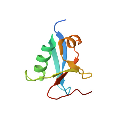
CIDE domains form functionally important higher-order assemblies for DNA fragmentation.

Choi, J.Y.Qiao, Q.Hong, S.H.Kim, C.M.Jeong, J.H.Kim, Y.G.Jung, Y.K.Wu, H.Park, H.H.

(2017) Proc Natl Acad Sci U S A 114: 7361-7366

  • DOI: https://doi.org/10.1073/pnas.1705949114
  • Primary Citation of Related Structures:  
    4D2K, 5XPC

  • PubMed Abstract: 

    Cell death-inducing DFF45-like effector (CIDE) domains, initially identified in apoptotic nucleases, form a family with diverse functions ranging from cell death to lipid homeostasis. Here we show that the CIDE domains of Drosophila and human apoptotic nucleases Drep2, Drep4, and DFF40 all form head-to-tail helical filaments. Opposing positively and negatively charged interfaces mediate the helical structures, and mutations on these surfaces abolish nuclease activation for apoptotic DNA fragmentation. Conserved filamentous structures are observed in CIDE family members involved in lipid homeostasis, and mutations on the charged interfaces compromise lipid droplet fusion, suggesting that CIDE domains represent a scaffold for higher-order assembly in DNA fragmentation and other biological processes such as lipid homeostasis.


  • Organizational Affiliation

    School of Chemistry and Biochemistry and Graduate School of Biochemistry, Yeungnam University, Gyeongsan 712-749, South Korea.


Macromolecules
Find similar proteins by:  (by identity cutoff)  |  3D Structure
Entity ID: 1
MoleculeChains Sequence LengthOrganismDetailsImage
DREP2
A, B, C, D
87Drosophila melanogasterMutation(s): 0 
UniProt
Find proteins for Q7K304 (Drosophila melanogaster)
Explore Q7K304 
Go to UniProtKB:  Q7K304
Entity Groups  
Sequence Clusters30% Identity50% Identity70% Identity90% Identity95% Identity100% Identity
UniProt GroupQ7K304
Sequence Annotations
Expand
  • Reference Sequence
Experimental Data & Validation

Experimental Data

  • Method: X-RAY DIFFRACTION
  • Resolution: 2.30 Å
  • R-Value Free: 0.254 
  • R-Value Work: 0.216 
  • R-Value Observed: 0.220 
  • Space Group: P 21 21 21
Unit Cell:
Length ( Å )Angle ( ˚ )
a = 50.283α = 90
b = 88.708β = 90
c = 113.372γ = 90
Software Package:
Software NamePurpose
PHENIXrefinement
HKL-2000data reduction
SCALEPACKdata scaling
PHASERphasing

Structure Validation

View Full Validation Report


Entry History 

Deposition Data

Revision History  (Full details and data files)

  • Version 1.0: 2015-05-27
    Type: Initial release
  • Version 1.1: 2017-06-28
    Changes: Database references
  • Version 1.2: 2017-07-12
    Changes: Database references
  • Version 1.3: 2017-08-02
    Changes: Database references
  • Version 1.4: 2023-12-20
    Changes: Data collection, Database references, Other, Refinement description