CLASP2 Has Two Distinct TOG Domains That Contribute Differently to Microtubule Dynamics
Maki, T., Grimaldi, A.D., Fuchigami, S., Kaverina, I., Hayashi, I.(2015) J Mol Biol 427: 2379-2395
- PubMed: 26003921
- DOI: https://doi.org/10.1016/j.jmb.2015.05.012
- Primary Citation of Related Structures:
3WOY, 3WOZ - PubMed Abstract:
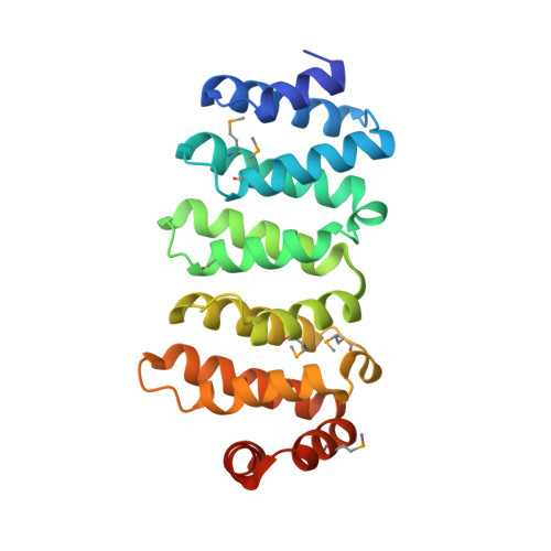
CLIP-associated proteins CLASPs are mammalian microtubule (MT) plus-end tracking proteins (+TIPs) that promote MT rescue in vivo. Their plus-end localization is dependent on other +TIPs, EB1 and CLIP-170, but in the leading edge of the cell, CLASPs display lattice-binding activity. MT association of CLASPs is suggested to be regulated by multiple TOG (tumor overexpressed gene) domains and by the serine-arginine (SR)-rich region, which contains binding sites for EB1. Here, we report the crystal structures of the two TOG domains of CLASP2. Both domains consist of six HEAT repeats, which are similar to the canonical paddle-like tubulin-binding TOG domains, but have arched conformations. The degrees and directions of curvature are different between the two TOG domains, implying that they have distinct roles in MT binding. Using biochemical, molecular modeling and cell biological analyses, we have investigated the interactions between the TOG domains and αβ-tubulin and found that each domain associates differently with αβ-tubulin. Our findings suggest that, by varying the degrees of domain curvature, the TOG domains may distinguish the structural conformation of the tubulin dimer, discriminate between different states of MT dynamic instability and thereby function differentially as stabilizers of MTs.
Organizational Affiliation:
Department of Medical Life Science, Yokohama City University, 1-7-29 Suehiro, Tsurumi, Yokohama, Kanagawa 230-0045, Japan.