3SI5

Kinetochore-BUBR1 kinase complex


Experimental Data Snapshot

  • Method: X-RAY DIFFRACTION
  • Resolution: 2.20 Å
  • R-Value Free: 0.247 
  • R-Value Work: 0.193 
  • R-Value Observed: 0.196 

wwPDB Validation   3D Report Full Report


This is version 1.2 of the entry. See complete history


Literature

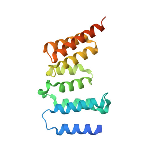
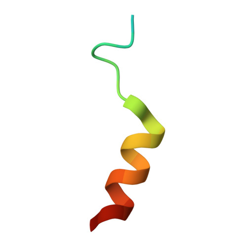
Structure of a Blinkin-BUBR1 Complex Reveals an Interaction Crucial for Kinetochore-Mitotic Checkpoint Regulation via an Unanticipated Binding Site.

Bolanos-Garcia, V.M.Lischetti, T.Matak-Vinkovic, D.Cota, E.Simpson, P.J.Chirgadze, D.Y.Spring, D.R.Robinson, C.V.Nilsson, J.Blundell, T.L.

(2011) Structure 19: 1691-1700

  • DOI: https://doi.org/10.1016/j.str.2011.09.017
  • Primary Citation of Related Structures:  
    3SI5

  • PubMed Abstract: 

    The maintenance of genomic stability relies on the spindle assembly checkpoint (SAC), which ensures accurate chromosome segregation by delaying the onset of anaphase until all chromosomes are properly bioriented and attached to the mitotic spindle. BUB1 and BUBR1 kinases are central for this process and by interacting with Blinkin, link the SAC with the kinetochore, the macromolecular assembly that connects microtubules with centromeric DNA. Here, we identify the Blinkin motif critical for interaction with BUBR1, define the stoichiometry and affinity of the interaction, and present a 2.2 Å resolution crystal structure of the complex. The structure defines an unanticipated BUBR1 region responsible for the interaction and reveals a novel Blinkin motif that undergoes a disorder-to-order transition upon ligand binding. We also show that substitution of several BUBR1 residues engaged in binding Blinkin leads to defects in the SAC, thus providing the first molecular details of the recognition mechanism underlying kinetochore-SAC signaling.


  • Organizational Affiliation

    Department of Biochemistry, University of Cambridge, Cambridge CB2 1GA, UK. victor@cryst.bioc.cam.ac.uk


Macromolecules
Find similar proteins by:  (by identity cutoff)  |  3D Structure
Entity ID: 1
MoleculeChains Sequence LengthOrganismDetailsImage
Mitotic checkpoint serine/threonine-protein kinase BUB1 beta
A, B
176Homo sapiensMutation(s): 0 
Gene Names: BUB1BBUBR1MAD3LSSK1
EC: 2.7.11.1
UniProt & NIH Common Fund Data Resources
Find proteins for O60566 (Homo sapiens)
Explore O60566 
Go to UniProtKB:  O60566
PHAROS:  O60566
GTEx:  ENSG00000156970 
Entity Groups  
Sequence Clusters30% Identity50% Identity70% Identity90% Identity95% Identity100% Identity
UniProt GroupO60566
Sequence Annotations
Expand
  • Reference Sequence

Find similar proteins by:  Sequence   |   3D Structure  

Entity ID: 2
MoleculeChains Sequence LengthOrganismDetailsImage
Protein CASC5C [auth X],
D [auth Y]
24Homo sapiensMutation(s): 0 
Gene Names: CASC5KIAA1570KNL1
UniProt & NIH Common Fund Data Resources
Find proteins for Q8NG31 (Homo sapiens)
Explore Q8NG31 
Go to UniProtKB:  Q8NG31
PHAROS:  Q8NG31
GTEx:  ENSG00000137812 
Entity Groups  
Sequence Clusters30% Identity50% Identity70% Identity90% Identity95% Identity100% Identity
UniProt GroupQ8NG31
Sequence Annotations
Expand
  • Reference Sequence
Experimental Data & Validation

Experimental Data

  • Method: X-RAY DIFFRACTION
  • Resolution: 2.20 Å
  • R-Value Free: 0.247 
  • R-Value Work: 0.193 
  • R-Value Observed: 0.196 
  • Space Group: C 1 2 1
Unit Cell:
Length ( Å )Angle ( ˚ )
a = 124.398α = 90
b = 40.143β = 91.38
c = 75.391γ = 90
Software Package:
Software NamePurpose
PROTEUM2data collection
AMoREphasing
REFMACrefinement
SAINTdata reduction
SADABSdata scaling

Structure Validation

View Full Validation Report


Entry History 

Deposition Data

Revision History  (Full details and data files)

  • Version 1.0: 2011-10-26
    Type: Initial release
  • Version 1.1: 2011-11-30
    Changes: Database references
  • Version 1.2: 2023-09-13
    Changes: Data collection, Database references, Refinement description