3OQT

Crystal structure of Rv1498A protein from mycobacterium tuberculosis


Experimental Data Snapshot

  • Method: X-RAY DIFFRACTION
  • Resolution: 2.88 Å
  • R-Value Free: 0.283 
  • R-Value Work: 0.252 
  • R-Value Observed: 0.254 

wwPDB Validation   3D Report Full Report


This is version 1.1 of the entry. See complete history


Literature

Structural and biophysical characterization of Mycobacterium tuberculosis dodecin Rv1498A.

Liu, F.Xiong, J.Kumar, S.Yang, C.Ge, S.Li, S.Xia, N.Swaminathan, K.

(2011) J Struct Biol 175: 31-38

  • DOI: https://doi.org/10.1016/j.jsb.2011.04.013
  • Primary Citation of Related Structures:  
    3OQT

  • PubMed Abstract: 

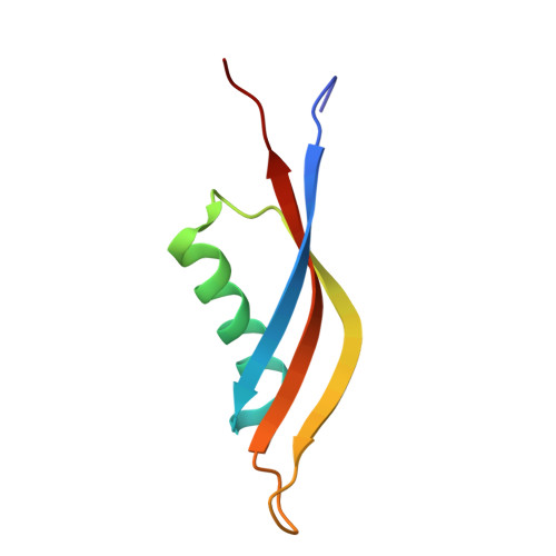
    Dodecins (assembly of twelve monomers) are the smallest known flavoprotein with only 65-73 amino acids and are involved in binding and storage of flavins in archaea. Here we report the crystal structure of Rv1498A, a Mycobacterium tuberculosis dodecin. This bacterial dodecin structure is similar to that of other reported dodecins. Each monomer has a 3 stranded β-sheet and an α-helix perpendicular to it. This protein has polyextreme (halophilic and thermophilic) properties. Interestingly, positively and negatively charged residues aggregate separately and do not seem to contribute to thermophilic and halophilic stability. We have examined the interactions that stabilize the Rv1498A dodecamer by preparing selected point mutants that break salt bridges and hydrophobic contacts, thereby leading to collapse of the assembly.


  • Organizational Affiliation

    Department of Biological Sciences, National University of Singapore, Singapore 117543, Singapore.


Macromolecules
Find similar proteins by:  (by identity cutoff)  |  3D Structure
Entity ID: 1
MoleculeChains Sequence LengthOrganismDetailsImage
Rv1498A PROTEIN
A, B, C, D, E
A, B, C, D, E, F, G, H, I, J, K, L, M, N, O, P
70Mycobacterium tuberculosisMutation(s): 0 
Gene Names: MT1547Rv1498.1Rv1498A
UniProt
Find proteins for Q8VK10 (Mycobacterium tuberculosis (strain CDC 1551 / Oshkosh))
Explore Q8VK10 
Go to UniProtKB:  Q8VK10
Entity Groups  
Sequence Clusters30% Identity50% Identity70% Identity90% Identity95% Identity100% Identity
UniProt GroupQ8VK10
Sequence Annotations
Expand
  • Reference Sequence
Small Molecules
Ligands 2 Unique
IDChains Name / Formula / InChI Key2D Diagram3D Interactions
CL
Query on CL

Download Ideal Coordinates CCD File 
AA [auth O]
BA [auth P]
Q [auth A]
R [auth C]
T [auth E]
AA [auth O],
BA [auth P],
Q [auth A],
R [auth C],
T [auth E],
U [auth H],
X [auth K],
Y [auth L]
CHLORIDE ION
Cl
VEXZGXHMUGYJMC-UHFFFAOYSA-M
NA
Query on NA

Download Ideal Coordinates CCD File 
CA [auth P],
S [auth C],
V [auth H],
W [auth I],
Z [auth L]
SODIUM ION
Na
FKNQFGJONOIPTF-UHFFFAOYSA-N
Experimental Data & Validation

Experimental Data

  • Method: X-RAY DIFFRACTION
  • Resolution: 2.88 Å
  • R-Value Free: 0.283 
  • R-Value Work: 0.252 
  • R-Value Observed: 0.254 
  • Space Group: P 21 3
Unit Cell:
Length ( Å )Angle ( ˚ )
a = 143.946α = 90
b = 143.946β = 90
c = 143.946γ = 90
Software Package:
Software NamePurpose
MOLREPphasing
PHASERphasing
REFMACrefinement
HKL-2000data reduction
HKL-2000data scaling

Structure Validation

View Full Validation Report


Entry History 

Deposition Data

Revision History  (Full details and data files)

  • Version 1.0: 2011-07-20
    Type: Initial release
  • Version 1.1: 2023-11-01
    Changes: Data collection, Database references, Derived calculations, Refinement description