3O7Q

Crystal structure of a Major Facilitator Superfamily (MFS) transporter, FucP, in the outward conformation


Experimental Data Snapshot

  • Method: X-RAY DIFFRACTION
  • Resolution: 3.14 Å
  • R-Value Free: 0.273 
  • R-Value Work: 0.215 
  • R-Value Observed: 0.218 

wwPDB Validation   3D Report Full Report


Ligand Structure Quality Assessment 


This is version 1.4 of the entry. See complete history


Literature

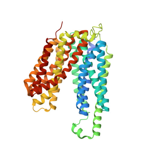
Structure of a fucose transporter in an outward-open conformation

Dang, S.Y.Sun, L.F.Huang, Y.Lu, F.Liu, Y.Gong, H.Wang, J.Yan, N.

(2010) Nature 467: 734-738

  • DOI: https://doi.org/10.1038/nature09406
  • Primary Citation of Related Structures:  
    3O7P, 3O7Q

  • PubMed Abstract: 

    The major facilitator superfamily (MFS) transporters are an ancient and widespread family of secondary active transporters. In Escherichia coli, the uptake of l-fucose, a source of carbon for microorganisms, is mediated by an MFS proton symporter, FucP. Despite intensive study of the MFS transporters, atomic structure information is only available on three proteins and the outward-open conformation has yet to be captured. Here we report the crystal structure of FucP at 3.1 Å resolution, which shows that it contains an outward-open, amphipathic cavity. The similarly folded amino and carboxyl domains of FucP have contrasting surface features along the transport path, with negative electrostatic potential on the N domain and hydrophobic surface on the C domain. FucP only contains two acidic residues along the transport path, Asp 46 and Glu 135, which can undergo cycles of protonation and deprotonation. Their essential role in active transport is supported by both in vivo and in vitro experiments. Structure-based biochemical analyses provide insights into energy coupling, substrate recognition and the transport mechanism of FucP.


  • Organizational Affiliation

    State Key Laboratory of Bio-membrane and Membrane Biotechnology, Center for Structural Biology, School of Life Sciences and School of Medicine, Tsinghua University, Beijing 100084, China.


Macromolecules
Find similar proteins by:  (by identity cutoff)  |  3D Structure
Entity ID: 1
MoleculeChains Sequence LengthOrganismDetailsImage
L-fucose-proton symporter438Escherichia coli K-12Mutation(s): 0 
Gene Names: fucPb2801JW2772
Membrane Entity: Yes 
UniProt
Find proteins for P11551 (Escherichia coli (strain K12))
Explore P11551 
Go to UniProtKB:  P11551
Entity Groups  
Sequence Clusters30% Identity50% Identity70% Identity90% Identity95% Identity100% Identity
UniProt GroupP11551
Sequence Annotations
Expand
  • Reference Sequence
Small Molecules
Ligands 1 Unique
IDChains Name / Formula / InChI Key2D Diagram3D Interactions
BNG
Query on BNG

Download Ideal Coordinates CCD File 
B [auth A]nonyl beta-D-glucopyranoside
C15 H30 O6
QFAPUKLCALRPLH-UXXRCYHCSA-N
Experimental Data & Validation

Experimental Data

  • Method: X-RAY DIFFRACTION
  • Resolution: 3.14 Å
  • R-Value Free: 0.273 
  • R-Value Work: 0.215 
  • R-Value Observed: 0.218 
  • Space Group: P 32 2 1
Unit Cell:
Length ( Å )Angle ( ˚ )
a = 119.46α = 90
b = 119.46β = 90
c = 98.36γ = 120
Software Package:
Software NamePurpose
BSSdata collection
PHASERphasing
PHENIXrefinement
DENZOdata reduction
SCALEPACKdata scaling

Structure Validation

View Full Validation Report


Ligand Structure Quality Assessment 


Entry History 

Deposition Data

Revision History  (Full details and data files)

  • Version 1.0: 2010-09-15
    Type: Initial release
  • Version 1.1: 2011-07-13
    Changes: Version format compliance
  • Version 1.2: 2014-03-26
    Changes: Database references
  • Version 1.3: 2020-07-29
    Type: Remediation
    Reason: Carbohydrate remediation
    Changes: Data collection, Derived calculations, Structure summary
  • Version 1.4: 2024-03-20
    Changes: Data collection, Database references, Structure summary