3LLA

Crystal Structure of the Alpha-kinase Domain of Myosin Heavy Chain Kinase A Complex with AMPPCP


Experimental Data Snapshot

  • Method: X-RAY DIFFRACTION
  • Resolution: 2.11 Å
  • R-Value Free: 0.263 
  • R-Value Work: 0.213 
  • R-Value Observed: 0.215 

wwPDB Validation   3D Report Full Report


Ligand Structure Quality Assessment 


This is version 1.2 of the entry. See complete history


Literature

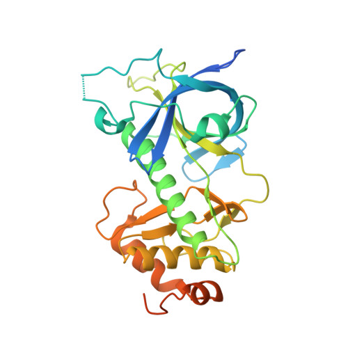
Crystal Structure of the {alpha}-Kinase Domain of Dictyostelium Myosin Heavy Chain Kinase A.

Ye, Q.Crawley, S.W.Yang, Y.Cote, G.P.Jia, Z.

(2010) Sci Signal 3: ra17-ra17

  • DOI: https://doi.org/10.1126/scisignal.2000525
  • Primary Citation of Related Structures:  
    3LKM, 3LLA, 3LMH, 3LMI

  • PubMed Abstract: 

    Dictyostelium discoideum myosin II heavy chain kinase A (MHCK A) disrupts the assembly and cellular activity of bipolar filaments of myosin II by phosphorylating sites within its alpha-helical, coiled-coil tail. MHCK A is a member of the atypical alpha-kinase family of serine and threonine protein kinases and displays no sequence homology to typical eukaryotic protein kinases. We report the crystal structure of the alpha-kinase domain (A-CAT) of MHCK A. When crystallized in the presence of adenosine triphosphate (ATP), A-CAT contained adenosine monophosphate (AMP) at the active site. However, when crystallized in the presence of ATP and a peptide substrate, which does not appear in the structure, adenosine diphosphate (ADP) was found at the active site and an invariant aspartic acid residue (Asp(766)) at the active site was phosphorylated. The aspartylphosphate group was exposed to the solvent within an active-site pocket that might function as a docking site for substrates. Access to the aspartylphosphate was regulated by a conformational switch in a loop that bound to a magnesium ion (Mg(2+)), providing a mechanism that allows alpha-kinases to sense and respond to local changes in Mg(2+).


  • Organizational Affiliation

    Department of Biochemistry, Queen's University, Kingston, Ontario, Canada K7L 3N6.


Macromolecules
Find similar proteins by:  (by identity cutoff)  |  3D Structure
Entity ID: 1
MoleculeChains Sequence LengthOrganismDetailsImage
Myosin heavy chain kinase A
A, B
307Dictyostelium discoideumMutation(s): 0 
Gene Names: DDB_G0291231mhckAmhkA
EC: 2.7.11.7
UniProt
Find proteins for P42527 (Dictyostelium discoideum)
Explore P42527 
Go to UniProtKB:  P42527
Entity Groups  
Sequence Clusters30% Identity50% Identity70% Identity90% Identity95% Identity100% Identity
UniProt GroupP42527
Sequence Annotations
Expand
  • Reference Sequence
Experimental Data & Validation

Experimental Data

  • Method: X-RAY DIFFRACTION
  • Resolution: 2.11 Å
  • R-Value Free: 0.263 
  • R-Value Work: 0.213 
  • R-Value Observed: 0.215 
  • Space Group: P 21 21 2
Unit Cell:
Length ( Å )Angle ( ˚ )
a = 83.075α = 90
b = 109.808β = 90
c = 79.626γ = 90
Software Package:
Software NamePurpose
ADSCdata collection
PHASERphasing
REFMACrefinement
HKL-2000data reduction
HKL-2000data scaling

Structure Validation

View Full Validation Report


Ligand Structure Quality Assessment 


Entry History 

Deposition Data

  • Released Date: 2010-03-16 
  • Deposition Author(s): Ye, Q., Jia, Z.

Revision History  (Full details and data files)

  • Version 1.0: 2010-03-16
    Type: Initial release
  • Version 1.1: 2011-07-13
    Changes: Version format compliance
  • Version 1.2: 2023-09-06
    Changes: Data collection, Database references, Derived calculations, Refinement description