3KYI

Crystal structure of the phosphorylated P1 domain of CheA3 in complex with CheY6 from R. sphaeroides


Experimental Data Snapshot

  • Method: X-RAY DIFFRACTION
  • Resolution: 2.80 Å
  • R-Value Free: 0.284 
  • R-Value Work: 0.241 
  • R-Value Observed: 0.243 

wwPDB Validation   3D Report Full Report


This is version 1.5 of the entry. See complete history


Literature

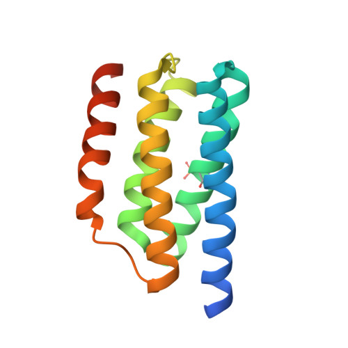
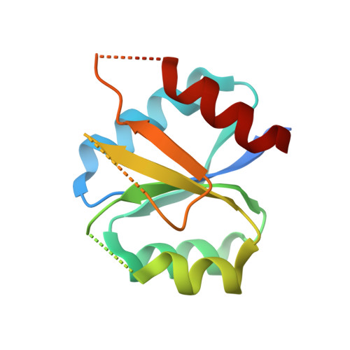
Using structural information to change the phosphotransfer specificity of a two-component chemotaxis signalling complex

Bell, C.H.Porter, S.L.Strawson, A.Stuart, D.I.Armitage, J.P.

(2010) PLoS Biol 8: e1000306-e1000306

  • DOI: https://doi.org/10.1371/journal.pbio.1000306
  • Primary Citation of Related Structures:  
    3KYI, 3KYJ

  • PubMed Abstract: 

    Two-component signal transduction pathways comprising histidine protein kinases (HPKs) and their response regulators (RRs) are widely used to control bacterial responses to environmental challenges. Some bacteria have over 150 different two-component pathways, and the specificity of the phosphotransfer reactions within these systems is tightly controlled to prevent unwanted crosstalk. One of the best understood two-component signalling pathways is the chemotaxis pathway. Here, we present the 1.40 A crystal structure of the histidine-containing phosphotransfer domain of the chemotaxis HPK, CheA(3), in complex with its cognate RR, CheY(6). A methionine finger on CheY(6) that nestles in a hydrophobic pocket in CheA(3) was shown to be important for the interaction and was found to only occur in the cognate RRs of CheA(3), CheY(6), and CheB(2). Site-directed mutagenesis of this methionine in combination with two adjacent residues abolished binding, as shown by surface plasmon resonance studies, and phosphotransfer from CheA(3)-P to CheY(6). Introduction of this methionine and an adjacent alanine residue into a range of noncognate CheYs, dramatically changed their specificity, allowing protein interaction and rapid phosphotransfer from CheA(3)-P. The structure presented here has allowed us to identify specificity determinants for the CheA-CheY interaction and subsequently to successfully reengineer phosphotransfer signalling. In summary, our results provide valuable insight into how cells mediate specificity in one of the most abundant signalling pathways in biology, two-component signal transduction.


  • Organizational Affiliation

    Oxford Centre for Integrative Systems Biology, Department of Biochemistry, University of Oxford, Oxford, United Kingdom.


Macromolecules
Find similar proteins by:  (by identity cutoff)  |  3D Structure
Entity ID: 1
MoleculeChains Sequence LengthOrganismDetailsImage
Putative histidine protein kinase144Cereibacter sphaeroidesMutation(s): 0 
Gene Names: cheA3
UniProt
Find proteins for Q8KLS0 (Cereibacter sphaeroides)
Explore Q8KLS0 
Go to UniProtKB:  Q8KLS0
Entity Groups  
Sequence Clusters30% Identity50% Identity70% Identity90% Identity95% Identity100% Identity
UniProt GroupQ8KLS0
Sequence Annotations
Expand
  • Reference Sequence
Find similar proteins by:  (by identity cutoff)  |  3D Structure
Entity ID: 2
MoleculeChains Sequence LengthOrganismDetailsImage
CheY6 protein145Cereibacter sphaeroidesMutation(s): 2 
Gene Names: cheY6
UniProt
Find proteins for Q8KLS1 (Cereibacter sphaeroides)
Explore Q8KLS1 
Go to UniProtKB:  Q8KLS1
Entity Groups  
Sequence Clusters30% Identity50% Identity70% Identity90% Identity95% Identity100% Identity
UniProt GroupQ8KLS1
Sequence Annotations
Expand
  • Reference Sequence
Small Molecules
Modified Residues  1 Unique
IDChains TypeFormula2D DiagramParent
NEP
Query on NEP
A
L-PEPTIDE LINKINGC6 H10 N3 O5 PHIS
Experimental Data & Validation

Experimental Data

  • Method: X-RAY DIFFRACTION
  • Resolution: 2.80 Å
  • R-Value Free: 0.284 
  • R-Value Work: 0.241 
  • R-Value Observed: 0.243 
  • Space Group: P 1
Unit Cell:
Length ( Å )Angle ( ˚ )
a = 33.17α = 78.81
b = 43.433β = 86.57
c = 48.647γ = 80.87
Software Package:
Software NamePurpose
DENZOdata reduction
SCALEPACKdata scaling
TNTrefinement
PDB_EXTRACTdata extraction
BUSTERrefinement

Structure Validation

View Full Validation Report


Entry History 

Deposition Data

Revision History  (Full details and data files)

  • Version 1.0: 2010-02-16
    Type: Initial release
  • Version 1.1: 2011-07-13
    Changes: Version format compliance
  • Version 1.2: 2016-06-15
    Changes: Data collection
  • Version 1.3: 2017-11-01
    Changes: Refinement description
  • Version 1.4: 2021-11-10
    Changes: Database references, Derived calculations
  • Version 1.5: 2023-11-01
    Changes: Data collection, Refinement description