3DL8

Structure of the complex of aquifex aeolicus SecYEG and bacillus subtilis SecA


Experimental Data Snapshot

  • Method: X-RAY DIFFRACTION
  • Resolution: 7.50 Å
  • R-Value Free: 0.390 
  • R-Value Work: 0.365 
  • R-Value Observed: 0.367 

wwPDB Validation   3D Report Full Report


This is version 1.3 of the entry. See complete history


Literature

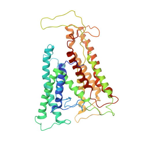
Structure of a complex of the ATPase SecA and the protein-translocation channel.

Zimmer, J.Nam, Y.Rapoport, T.A.

(2008) Nature 455: 936-943

  • DOI: https://doi.org/10.1038/nature07335
  • Primary Citation of Related Structures:  
    3DIN, 3DL8

  • PubMed Abstract: 

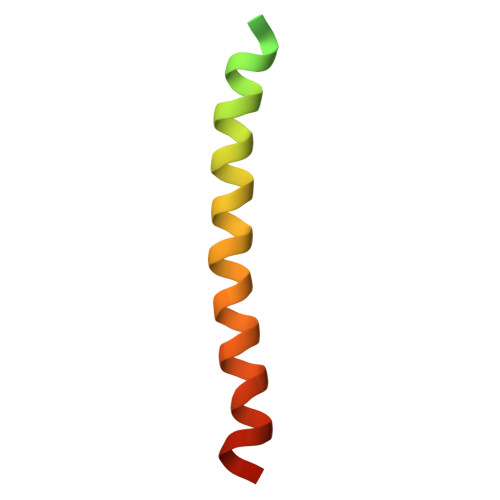
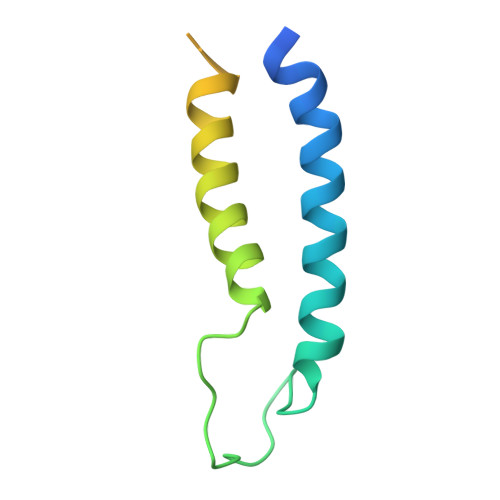
    Most proteins are secreted from bacteria by the interaction of the cytoplasmic SecA ATPase with a membrane channel, formed by the heterotrimeric SecY complex. Here we report the crystal structure of SecA bound to the SecY complex, with a maximum resolution of 4.5 ångström (A), obtained for components from Thermotoga maritima. One copy of SecA in an intermediate state of ATP hydrolysis is bound to one molecule of the SecY complex. Both partners undergo important conformational changes on interaction. The polypeptide-cross-linking domain of SecA makes a large conformational change that could capture the translocation substrate in a 'clamp'. Polypeptide movement through the SecY channel could be achieved by the motion of a 'two-helix finger' of SecA inside the cytoplasmic funnel of SecY, and by the coordinated tightening and widening of SecA's clamp above the SecY pore. SecA binding generates a 'window' at the lateral gate of the SecY channel and it displaces the plug domain, preparing the channel for signal sequence binding and channel opening.


  • Organizational Affiliation

    Howard Hughes Medical Institute and Department of Cell Biology, Harvard Medical School, 240 Longwood Avenue, Boston, Massachusetts 02115, USA.


Macromolecules
Find similar proteins by:  (by identity cutoff)  |  3D Structure
Entity ID: 1
MoleculeChains Sequence LengthOrganismDetailsImage
Protein translocase subunit secA
A, B
779Bacillus subtilisMutation(s): 0 
Gene Names: secAdiv+BSU35300
Membrane Entity: Yes 
UniProt
Find proteins for P28366 (Bacillus subtilis (strain 168))
Explore P28366 
Go to UniProtKB:  P28366
Entity Groups  
Sequence Clusters30% Identity50% Identity70% Identity90% Identity95% Identity100% Identity
UniProt GroupP28366
Sequence Annotations
Expand
  • Reference Sequence
Find similar proteins by:  (by identity cutoff)  |  3D Structure
Entity ID: 2
MoleculeChains Sequence LengthOrganismDetailsImage
Preprotein translocase subunit secYC [auth G],
D [auth H]
429Aquifex aeolicusMutation(s): 0 
Gene Names: secYaq_079
Membrane Entity: Yes 
UniProt
Find proteins for O66491 (Aquifex aeolicus (strain VF5))
Explore O66491 
Go to UniProtKB:  O66491
Entity Groups  
Sequence Clusters30% Identity50% Identity70% Identity90% Identity95% Identity100% Identity
UniProt GroupO66491
Sequence Annotations
Expand
  • Reference Sequence
Find similar proteins by:  (by identity cutoff)  |  3D Structure
Entity ID: 3
MoleculeChains Sequence LengthOrganismDetailsImage
SecEE [auth C],
F [auth D]
65Aquifex aeolicusMutation(s): 0 
Membrane Entity: Yes 
UniProt
Find proteins for D0VWU4 (Aquifex aeolicus (strain VF5))
Explore D0VWU4 
Go to UniProtKB:  D0VWU4
Entity Groups  
Sequence Clusters30% Identity50% Identity70% Identity90% Identity95% Identity100% Identity
UniProt GroupD0VWU4
Sequence Annotations
Expand
  • Reference Sequence
Find similar proteins by:  (by identity cutoff)  |  3D Structure
Entity ID: 4
MoleculeChains Sequence LengthOrganismDetailsImage
Protein-export membrane protein secGG [auth E],
H [auth F]
107Aquifex aeolicusMutation(s): 0 
Gene Names: secGaq_098
Membrane Entity: Yes 
UniProt
Find proteins for O66505 (Aquifex aeolicus (strain VF5))
Explore O66505 
Go to UniProtKB:  O66505
Entity Groups  
Sequence Clusters30% Identity50% Identity70% Identity90% Identity95% Identity100% Identity
UniProt GroupO66505
Sequence Annotations
Expand
  • Reference Sequence
Experimental Data & Validation

Experimental Data

  • Method: X-RAY DIFFRACTION
  • Resolution: 7.50 Å
  • R-Value Free: 0.390 
  • R-Value Work: 0.365 
  • R-Value Observed: 0.367 
  • Space Group: P 21 21 21
Unit Cell:
Length ( Å )Angle ( ˚ )
a = 146.359α = 90
b = 167.974β = 90
c = 187.722γ = 90
Software Package:
Software NamePurpose
CNSrefinement
PDB_EXTRACTdata extraction
HKL-2000data collection
HKL-2000data reduction
HKL-2000data scaling
PHASERphasing

Structure Validation

View Full Validation Report


Entry History 

Deposition Data

Revision History  (Full details and data files)

  • Version 1.0: 2008-12-02
    Type: Initial release
  • Version 1.1: 2011-07-13
    Changes: Version format compliance
  • Version 1.2: 2013-09-25
    Changes: Derived calculations
  • Version 1.3: 2024-02-21
    Changes: Data collection, Database references