3DL6

Crystal Structure of the A287F/S290G Active Site Mutant of TS-DHFR from Cryptosporidium hominis


Experimental Data Snapshot

  • Method: X-RAY DIFFRACTION
  • Resolution: 3.25 Å
  • R-Value Free: 0.276 
  • R-Value Work: 0.225 
  • R-Value Observed: 0.225 

wwPDB Validation   3D Report Full Report


Ligand Structure Quality Assessment 


This is version 1.3 of the entry. See complete history


Literature

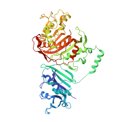
Explaining an unusually fast parasitic enzyme: folate tail-binding residues dictate substrate positioning and catalysis in Cryptosporidium hominis thymidylate synthase.

Martucci, W.E.Vargo, M.A.Anderson, K.S.

(2008) Biochemistry 47: 8902-8911

  • DOI: https://doi.org/10.1021/bi800466z
  • Primary Citation of Related Structures:  
    3DL5, 3DL6

  • PubMed Abstract: 

    The essential enzyme TS-DHFR from Cryptosporidium hominis undergoes an unusually rapid rate of catalysis at the conserved TS domain, facilitated by two nonconserved residues, Ala287 and Ser290, in the folate tail-binding region. Mutation of these two residues to their conserved counterparts drastically affects multiple steps of the TS catalytic cycle. We have determined the crystal structures of all three mutants (A287F, S290G, and A287F/S290G) in complex with active site ligands dUMP and CB3717. The structural data show two effects of the mutations: an increased distance between the ligands in the active site and increased flexibility of the folate ligand in the partially open enzyme state that precedes conformational change to the active catalytic state. The latter effect is able to be rescued by the mutants containing the A287F mutation. In addition, the conserved water network of TS is altered in each of the mutants. The structural results point to a role of the folate tail-binding residues in closely positioning ChTS ligands and restricting ligand flexibility in the partially open state to allow for a rapid transition to the active closed state and enhanced rate of catalysis. These results provide an explanation on how folate tail-binding residues at one end of the active site affect long-range interactions throughout the TS active site and validate these residues as targets for species-specific drug design.


  • Organizational Affiliation

    Department of Molecular Biophysics and Biochemistry, Yale University School of Medicine, 333 Cedar Street, New Haven, Connecticut 06520, USA.


Macromolecules
Find similar proteins by:  (by identity cutoff)  |  3D Structure
Entity ID: 1
MoleculeChains Sequence LengthOrganismDetailsImage
Dihydrofolate reductase, DHFR
A, B, C, D, E
521Cryptosporidium hominisMutation(s): 2 
Gene Names: Chro.40506
EC: 1.5.1.3
Entity Groups  
Sequence Clusters30% Identity50% Identity70% Identity90% Identity95% Identity100% Identity
Sequence Annotations
Expand
  • Reference Sequence
Small Molecules
Ligands 4 Unique
IDChains Name / Formula / InChI Key2D Diagram3D Interactions
NDP
Query on NDP

Download Ideal Coordinates CCD File 
I [auth A],
M [auth B],
Q [auth C],
U [auth D],
Y [auth E]
NADPH DIHYDRO-NICOTINAMIDE-ADENINE-DINUCLEOTIDE PHOSPHATE
C21 H30 N7 O17 P3
ACFIXJIJDZMPPO-NNYOXOHSSA-N
CB3
Query on CB3

Download Ideal Coordinates CCD File 
G [auth A],
K [auth B],
O [auth C],
S [auth D],
W [auth E]
10-PROPARGYL-5,8-DIDEAZAFOLIC ACID
C24 H23 N5 O6
LTKHPMDRMUCUEB-IBGZPJMESA-N
DHF
Query on DHF

Download Ideal Coordinates CCD File 
H [auth A],
L [auth B],
P [auth C],
T [auth D],
X [auth E]
DIHYDROFOLIC ACID
C19 H21 N7 O6
OZRNSSUDZOLUSN-LBPRGKRZSA-N
UMP
Query on UMP

Download Ideal Coordinates CCD File 
F [auth A],
J [auth B],
N [auth C],
R [auth D],
V [auth E]
2'-DEOXYURIDINE 5'-MONOPHOSPHATE
C9 H13 N2 O8 P
JSRLJPSBLDHEIO-SHYZEUOFSA-N
Experimental Data & Validation

Experimental Data

  • Method: X-RAY DIFFRACTION
  • Resolution: 3.25 Å
  • R-Value Free: 0.276 
  • R-Value Work: 0.225 
  • R-Value Observed: 0.225 
  • Space Group: C 1 2 1
Unit Cell:
Length ( Å )Angle ( ˚ )
a = 214.906α = 90
b = 116.921β = 95.94
c = 220.948γ = 90
Software Package:
Software NamePurpose
CBASSdata collection
AMoREphasing
CNSrefinement
HKL-2000data reduction
HKL-2000data scaling

Structure Validation

View Full Validation Report


Ligand Structure Quality Assessment 


Entry History 

Deposition Data

Revision History  (Full details and data files)

  • Version 1.0: 2008-08-19
    Type: Initial release
  • Version 1.1: 2011-07-13
    Changes: Version format compliance
  • Version 1.2: 2021-10-20
    Changes: Database references, Derived calculations
  • Version 1.3: 2023-08-30
    Changes: Data collection, Refinement description