2Y6T

Molecular Recognition of Chymotrypsin by the Serine Protease Inhibitor Ecotin from Yersinia pestis


Experimental Data Snapshot

  • Method: X-RAY DIFFRACTION
  • Resolution: 2.74 Å
  • R-Value Free: 0.314 
  • R-Value Work: 0.244 
  • R-Value Observed: 0.247 

wwPDB Validation   3D Report Full Report


This is version 1.3 of the entry. See complete history


Literature

Molecular Recognition of Chymotrypsin by the Serine Protease Inhibitor Ecotin from Yersinia Pestis.

Clark, E.A.Walker, N.Ford, D.C.Cooper, I.A.Oyston, P.C.Acharya, K.R.

(2011) J Biol Chem 286: 24015

  • DOI: https://doi.org/10.1074/jbc.M111.225730
  • Primary Citation of Related Structures:  
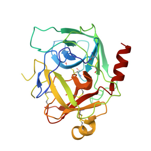
    2Y6T

  • PubMed Abstract: 

    Resistance to antibiotics is a problem not only in terms of healthcare but also biodefense. Engineering of resistance into a human pathogen could create an untreatable biothreat pathogen. One such pathogen is Yersinia pestis, the causative agent of plague. Previously, we have used a bioinformatic approach to identify proteins that may be suitable targets for antimicrobial therapy and in particular for the treatment of plague. The serine protease inhibitor ecotin was identified as one such target. We have carried out mutational analyses in the closely related Yersinia pseudotuberculosis, validating that the ecotin gene is a virulence-associated gene in this bacterium. Y. pestis ecotin inhibits chymotrypsin. Here, we present the structure of ecotin in complex with chymotrypsin to 2.74 Å resolution. The structure features a biologically relevant tetramer whereby an ecotin dimer binds to two chymotrypsin molecules, similar to what was observed in related serine protease inhibitor structures. However, the vast majority of the interactions in the present structure are distinctive, indicating that the broad specificity of the inhibitor for these proteases is based largely on its capacity to recognize features unique to each of them. These findings will have implications for the development of small ecotin inhibitors for therapeutic use.


  • Organizational Affiliation

    Department of Biology and Biochemistry, University of Bath, Claverton Down, Bath BA2 7AY, United Kingdom.


Macromolecules
Find similar proteins by:  (by identity cutoff)  |  3D Structure
Entity ID: 1
MoleculeChains Sequence LengthOrganismDetailsImage
CHYMOTRYPSINOGEN A
A, B, C, D
245Bos taurusMutation(s): 0 
EC: 3.4.21.1
UniProt
Find proteins for P00766 (Bos taurus)
Explore P00766 
Go to UniProtKB:  P00766
Entity Groups  
Sequence Clusters30% Identity50% Identity70% Identity90% Identity95% Identity100% Identity
UniProt GroupP00766
Sequence Annotations
Expand
  • Reference Sequence
Find similar proteins by:  (by identity cutoff)  |  3D Structure
Entity ID: 2
MoleculeChains Sequence LengthOrganismDetailsImage
ECOTIN
E, F, G, H
148Yersinia pseudotuberculosis YPIIIMutation(s): 0 
UniProt
Find proteins for B1JSA0 (Yersinia pseudotuberculosis serotype O:3 (strain YPIII))
Explore B1JSA0 
Go to UniProtKB:  B1JSA0
Entity Groups  
Sequence Clusters30% Identity50% Identity70% Identity90% Identity95% Identity100% Identity
UniProt GroupB1JSA0
Sequence Annotations
Expand
  • Reference Sequence
Experimental Data & Validation

Experimental Data

  • Method: X-RAY DIFFRACTION
  • Resolution: 2.74 Å
  • R-Value Free: 0.314 
  • R-Value Work: 0.244 
  • R-Value Observed: 0.247 
  • Space Group: P 1 21 1
Unit Cell:
Length ( Å )Angle ( ˚ )
a = 97.31α = 90
b = 48.278β = 103.96
c = 174.636γ = 90
Software Package:
Software NamePurpose
REFMACrefinement
HKL-2000data reduction
HKL-2000data scaling
PHASERphasing

Structure Validation

View Full Validation Report


Entry History 

Deposition Data

Revision History  (Full details and data files)

  • Version 1.0: 2011-04-20
    Type: Initial release
  • Version 1.1: 2013-11-27
    Changes: Source and taxonomy, Version format compliance
  • Version 1.2: 2017-03-29
    Changes: Source and taxonomy
  • Version 1.3: 2023-12-20
    Changes: Data collection, Database references, Derived calculations, Other, Refinement description