2XXN

Structure of the vIRF4-HAUSP TRAF domain complex


Experimental Data Snapshot

  • Method: X-RAY DIFFRACTION
  • Resolution: 1.60 Å
  • R-Value Free: 0.174 
  • R-Value Work: 0.158 
  • R-Value Observed: 0.159 

wwPDB Validation   3D Report Full Report


This is version 1.3 of the entry. See complete history


Literature

Bilateral Inhibition of Hausp Deubiquitinase by a Viral Interferon Regulatory Factor Protein

Lee, H.R.Choi, W.C.Lee, S.Hwang, J.Hwang, E.Guchhait, K.Haas, J.Toth, Z.Jeon, Y.H.Oh, T.K.Kim, M.H.Jung, J.U.

(2011) Nat Struct Mol Biol 18: 1336

  • DOI: https://doi.org/10.1038/nsmb.2142
  • Primary Citation of Related Structures:  
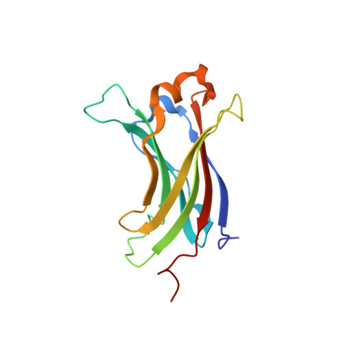
    2XXN

  • PubMed Abstract: 

    Herpesvirus-associated ubiquitin-specific protease (HAUSP) regulates the stability of p53 and the p53-binding protein MDM2, implicating HAUSP as a therapeutic target for tuning p53-mediated antitumor activity. Here we report the structural analysis of HAUSP with Kaposi's sarcoma-associated herpesvirus viral interferon (IFN) regulatory factor 4 (vIRF4) and the discovery of two vIRF4-derived peptides, vif1 and vif2, as potent and selective HAUSP antagonists. This analysis reveals a bilateral belt-type interaction that results in inhibition of HAUSP. The vif1 peptide binds the HAUSP TRAF domain, competitively blocking substrate binding, whereas the vif2 peptide binds both the HAUSP TRAF and catalytic domains, robustly suppressing its deubiquitination activity. Peptide treatments comprehensively blocked HAUSP, leading to p53-dependent cell-cycle arrest and apoptosis in culture and to tumor regression in xenograft mouse model. Thus, the virus has developed a unique strategy to target the HAUSP-MDM2-p53 pathway, and these virus-derived short peptides represent biologically active HAUSP antagonists.


  • Organizational Affiliation

    Department of Molecular Microbiology and Immunology, Keck School of Medicine, University of Southern California, Los Angeles, California, USA.


Macromolecules
Find similar proteins by:  (by identity cutoff)  |  3D Structure
Entity ID: 1
MoleculeChains Sequence LengthOrganismDetailsImage
UBIQUITIN CARBOXYL-TERMINAL HYDROLASE 7143Homo sapiensMutation(s): 0 
EC: 3.4.19.12
UniProt & NIH Common Fund Data Resources
Find proteins for Q93009 (Homo sapiens)
Explore Q93009 
Go to UniProtKB:  Q93009
PHAROS:  Q93009
GTEx:  ENSG00000187555 
Entity Groups  
Sequence Clusters30% Identity50% Identity70% Identity90% Identity95% Identity100% Identity
UniProt GroupQ93009
Sequence Annotations
Expand
  • Reference Sequence

Find similar proteins by:  Sequence   |   3D Structure  

Entity ID: 2
MoleculeChains Sequence LengthOrganismDetailsImage
K1015Human gammaherpesvirus 8Mutation(s): 0 
UniProt
Find proteins for Q2HR73 (Human herpesvirus 8 type P (isolate GK18))
Explore Q2HR73 
Go to UniProtKB:  Q2HR73
Entity Groups  
Sequence Clusters30% Identity50% Identity70% Identity90% Identity95% Identity100% Identity
UniProt GroupQ2HR73
Sequence Annotations
Expand
  • Reference Sequence
Experimental Data & Validation

Experimental Data

  • Method: X-RAY DIFFRACTION
  • Resolution: 1.60 Å
  • R-Value Free: 0.174 
  • R-Value Work: 0.158 
  • R-Value Observed: 0.159 
  • Space Group: P 32 2 1
Unit Cell:
Length ( Å )Angle ( ˚ )
a = 72.462α = 90
b = 72.462β = 90
c = 53.84γ = 120
Software Package:
Software NamePurpose
REFMACrefinement
MOLREPphasing

Structure Validation

View Full Validation Report


Entry History 

Deposition Data

Revision History  (Full details and data files)

  • Version 1.0: 2011-11-09
    Type: Initial release
  • Version 1.1: 2011-12-14
    Changes: Database references
  • Version 1.2: 2012-03-28
    Changes: Other
  • Version 1.3: 2023-12-20
    Changes: Data collection, Database references, Other, Refinement description