2VXG

Crystal structure of the conserved C-terminal region of Ge-1


Experimental Data Snapshot

  • Method: X-RAY DIFFRACTION
  • Resolution: 1.90 Å
  • R-Value Free: 0.254 
  • R-Value Work: 0.211 
  • R-Value Observed: 0.213 

wwPDB Validation   3D Report Full Report


This is version 1.2 of the entry. See complete history


Literature

The C-Terminal Region of Ge-1 Presents Conserved Structural Features Required for P-Body Localization.

Jinek, M.Eulalio, A.Lingel, A.Helms, S.Conti, E.Izaurralde, E.

(2008) RNA 14: 1991

  • DOI: https://doi.org/10.1261/rna.1222908
  • Primary Citation of Related Structures:  
    2VXG

  • PubMed Abstract: 

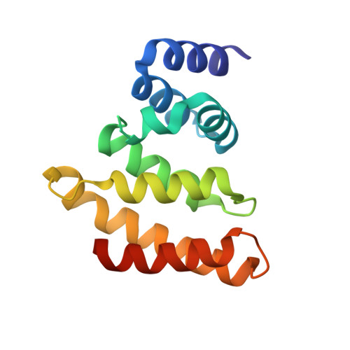
    The removal of the 5' cap structure by the DCP1-DCP2 decapping complex irreversibly commits eukaryotic mRNAs to degradation. In human cells, the interaction between DCP1 and DCP2 is bridged by the Ge-1 protein. Ge-1 contains an N-terminal WD40-repeat domain connected by a low-complexity region to a conserved C-terminal domain. It was reported that the C-terminal domain interacts with DCP2 and mediates Ge-1 oligomerization and P-body localization. To understand the molecular basis for these functions, we determined the three-dimensional crystal structure of the most conserved region of the Drosophila melanogaster Ge-1 C-terminal domain. The region adopts an all alpha-helical fold related to ARM- and HEAT-repeat proteins. Using structure-based mutants we identified an invariant surface residue affecting P-body localization. The conservation of critical surface and structural residues suggests that the C-terminal region adopts a similar fold with conserved functions in all members of the Ge-1 protein family.


  • Organizational Affiliation

    Department of Structural Cell Biology, Max Planck Institute of Biochemistry, D-82152 Martinsried, Germany.


Macromolecules
Find similar proteins by:  (by identity cutoff)  |  3D Structure
Entity ID: 1
MoleculeChains Sequence LengthOrganismDetailsImage
CG6181-PA, ISOFORM A
A, B
139Drosophila melanogasterMutation(s): 0 
UniProt
Find proteins for Q9VKK1 (Drosophila melanogaster)
Explore Q9VKK1 
Go to UniProtKB:  Q9VKK1
Entity Groups  
Sequence Clusters30% Identity50% Identity70% Identity90% Identity95% Identity100% Identity
UniProt GroupQ9VKK1
Sequence Annotations
Expand
  • Reference Sequence
Experimental Data & Validation

Experimental Data

  • Method: X-RAY DIFFRACTION
  • Resolution: 1.90 Å
  • R-Value Free: 0.254 
  • R-Value Work: 0.211 
  • R-Value Observed: 0.213 
  • Space Group: P 21 21 21
Unit Cell:
Length ( Å )Angle ( ˚ )
a = 38.24α = 90
b = 65.87β = 90
c = 103.78γ = 90
Software Package:
Software NamePurpose
REFMACrefinement
XDSdata reduction
XDSdata scaling
SHARPphasing

Structure Validation

View Full Validation Report


Entry History 

Deposition Data

Revision History  (Full details and data files)

  • Version 1.0: 2008-09-09
    Type: Initial release
  • Version 1.1: 2011-05-07
    Changes: Version format compliance
  • Version 1.2: 2011-07-13
    Changes: Version format compliance