2V1C

Crystal structure and mutational study of RecOR provide insight into its role in DNA repair


Experimental Data Snapshot

  • Method: X-RAY DIFFRACTION
  • Resolution: 3.80 Å
  • R-Value Free: 0.443 
  • R-Value Work: 0.459 
  • R-Value Observed: 0.458 

wwPDB Validation   3D Report Full Report


This is version 1.3 of the entry. See complete history


Literature

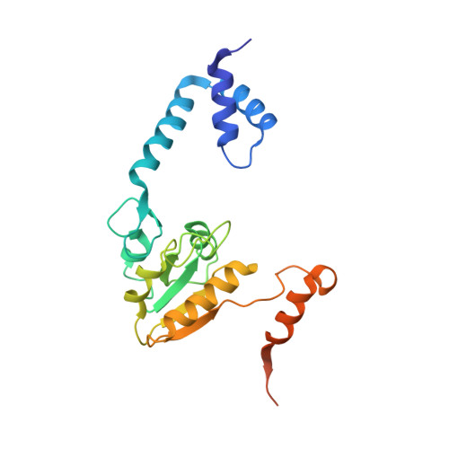
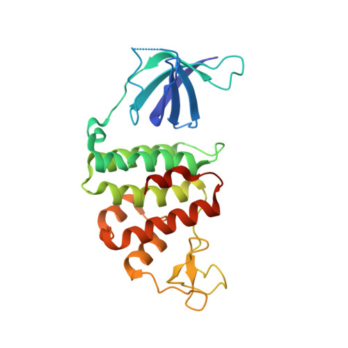
Crystal Structure and Mutational Study of Recor Provide Insight Into its Mode of DNA Binding.

Timmins, J.Leiros, I.Mcsweeney, S.

(2007) EMBO J 26: 3260

  • DOI: https://doi.org/10.1038/sj.emboj.7601760
  • Primary Citation of Related Structures:  
    2V1C

  • PubMed Abstract: 

    The crystal structure of the complex formed between Deinococcus radiodurans RecR and RecO (drRecOR) has been determined. In accordance with previous biochemical characterisation, the drRecOR complex displays a RecR:RecO molecular ratio of 2:1. The biologically relevant drRecOR entity consists of a heterohexamer in the form of two drRecO molecules positioned on either side of the tetrameric ring of drRecR, with their OB (oligonucleotide/oligosaccharide-binding) domains pointing towards the interior of the ring. Mutagenesis studies validated the protein-protein interactions observed in the crystal structure and allowed mapping of the residues in the drRecOR complex required for DNA binding. Furthermore, the preferred DNA substrate of drRecOR was identified as being 3'-overhanging DNA, as encountered at ssDNA-dsDNA junctions. Together these results suggest a possible mechanism for drRecOR recognition of stalled replication forks.


  • Organizational Affiliation

    Macromolecular Crystallography Group, European Synchrotron Radiation Facility, Grenoble-CEDEX 9, France.


Macromolecules
Find similar proteins by:  (by identity cutoff)  |  3D Structure
Entity ID: 1
MoleculeChains Sequence LengthOrganismDetailsImage
RECOMBINATION PROTEIN RECR
A, B
220Deinococcus radiodurans R1 = ATCC 13939 = DSM 20539Mutation(s): 0 
UniProt
Find proteins for Q9ZNA2 (Deinococcus radiodurans (strain ATCC 13939 / DSM 20539 / JCM 16871 / CCUG 27074 / LMG 4051 / NBRC 15346 / NCIMB 9279 / VKM B-1422 / R1))
Explore Q9ZNA2 
Go to UniProtKB:  Q9ZNA2
Entity Groups  
Sequence Clusters30% Identity50% Identity70% Identity90% Identity95% Identity100% Identity
UniProt GroupQ9ZNA2
Sequence Annotations
Expand
  • Reference Sequence
Find similar proteins by:  (by identity cutoff)  |  3D Structure
Entity ID: 2
MoleculeChains Sequence LengthOrganismDetailsImage
HYPOTHETICAL PROTEIN244Deinococcus radiodurans R1 = ATCC 13939 = DSM 20539Mutation(s): 0 
UniProt
Find proteins for Q9RW50 (Deinococcus radiodurans (strain ATCC 13939 / DSM 20539 / JCM 16871 / CCUG 27074 / LMG 4051 / NBRC 15346 / NCIMB 9279 / VKM B-1422 / R1))
Explore Q9RW50 
Go to UniProtKB:  Q9RW50
Entity Groups  
Sequence Clusters30% Identity50% Identity70% Identity90% Identity95% Identity100% Identity
UniProt GroupQ9RW50
Sequence Annotations
Expand
  • Reference Sequence
Experimental Data & Validation

Experimental Data

  • Method: X-RAY DIFFRACTION
  • Resolution: 3.80 Å
  • R-Value Free: 0.443 
  • R-Value Work: 0.459 
  • R-Value Observed: 0.458 
  • Space Group: C 1 2 1
Unit Cell:
Length ( Å )Angle ( ˚ )
a = 144α = 90
b = 83.2β = 106.9
c = 66.6γ = 90
Software Package:
Software NamePurpose
REFMACrefinement
XDSdata reduction
XDSdata scaling
PHASERphasing

Structure Validation

View Full Validation Report


Entry History 

Deposition Data

Revision History  (Full details and data files)

  • Version 1.0: 2007-07-03
    Type: Initial release
  • Version 1.1: 2011-05-08
    Changes: Version format compliance
  • Version 1.2: 2011-07-13
    Changes: Version format compliance
  • Version 1.3: 2023-12-13
    Changes: Data collection, Database references, Derived calculations, Other, Refinement description