2P32

Crystal structure of the C-terminal 10 kDa subdomain from C. elegans Hsp70


Experimental Data Snapshot

  • Method: X-RAY DIFFRACTION
  • Resolution: 3.20 Å
  • R-Value Free: 0.282 
  • R-Value Work: 0.268 

wwPDB Validation   3D Report Full Report


This is version 1.5 of the entry. See complete history


Literature

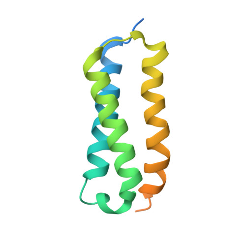
Crystal structure of the C-terminal three-helix bundle subdomain of C. elegans Hsp70.

Worrall, L.J.Walkinshaw, M.D.

(2007) Biochem Biophys Res Commun 357: 105-110

  • DOI: https://doi.org/10.1016/j.bbrc.2007.03.107
  • Primary Citation of Related Structures:  
    2P32

  • PubMed Abstract: 

    Hsp70 chaperones are composed of two domains; the 40 kDa N-terminal nucleotide-binding domain (NDB) and the 30 kDa C-terminal substrate-binding domain (SBD). Structures of the SBD from Escherichia coli homologues DnaK and HscA show it can be further divided into an 18 kDa beta-sandwich subdomain, which forms the hydrophobic binding pocket, and a 10 kDa C-terminal three-helix bundle that forms a lid over the binding pocket. Across prokaryotes and eukaryotes, the NBD and beta-sandwich subdomain are well conserved in both sequence and structure. The C-terminal subdomain is, however, more evolutionary variable and the only eukaryotic structure from rat Hsc70 revealed a diverged helix-loop-helix fold. We have solved the crystal structure of the C-terminal 10 kDa subdomain from Caenorhabditis elegans Hsp70 which forms a helical-bundle similar to the prokaryotic homologues. This provides the first confirmation of the structural conservation of this subdomain in eukaryotes. Comparison with the rat structure reveals a domain-swap dimerisation mechanism; however, the C. elegans subdomain exists exclusively as a monomer in solution in agreement with the hypothesis that regions out with the C-terminal subdomain are necessary for Hsp70 self-association.


  • Organizational Affiliation

    Centre for Translational and Chemical Biology, School of Biological Sciences, University of Edinburgh, Edinburgh EH9 3JR, UK.


Macromolecules
Find similar proteins by:  (by identity cutoff)  |  3D Structure
Entity ID: 1
MoleculeChains Sequence LengthOrganismDetailsImage
Heat shock 70 kDa protein A
A, B, C, D, E
A, B, C, D, E, F
120Caenorhabditis elegansMutation(s): 0 
Gene Names: hsp-1hsp70a
UniProt
Find proteins for P09446 (Caenorhabditis elegans)
Explore P09446 
Go to UniProtKB:  P09446
Entity Groups  
Sequence Clusters30% Identity50% Identity70% Identity90% Identity95% Identity100% Identity
UniProt GroupP09446
Sequence Annotations
Expand
  • Reference Sequence
Experimental Data & Validation

Experimental Data

  • Method: X-RAY DIFFRACTION
  • Resolution: 3.20 Å
  • R-Value Free: 0.282 
  • R-Value Work: 0.268 
  • Space Group: P 42 21 2
Unit Cell:
Length ( Å )Angle ( ˚ )
a = 138.927α = 90
b = 138.927β = 90
c = 100.704γ = 90
Software Package:
Software NamePurpose
SCALAdata scaling
REFMACrefinement
PDB_EXTRACTdata extraction
MAR345data collection
MOSFLMdata reduction
CCP4data scaling
PHASERphasing

Structure Validation

View Full Validation Report


Entry History 

Deposition Data

Revision History  (Full details and data files)

  • Version 1.0: 2007-04-17
    Type: Initial release
  • Version 1.1: 2008-05-01
    Changes: Version format compliance
  • Version 1.2: 2011-07-13
    Changes: Advisory, Derived calculations, Version format compliance
  • Version 1.3: 2017-10-18
    Changes: Refinement description
  • Version 1.4: 2024-02-21
    Changes: Data collection, Database references, Derived calculations, Refinement description
  • Version 1.5: 2024-04-03
    Changes: Refinement description