Solution Structure and Molecular Determinants of Hemoglobin Binding of the First NEAT Domain of IsdB in Staphylococcus aureus.
Fonner, B.A., Tripet, B.P., Eilers, B.J., Stanisich, J., Sullivan-Springhetti, R.K., Moore, R., Liu, M., Lei, B., Copie, V.(2014) Biochemistry 53: 3922-3933
- PubMed: 24871270
- DOI: https://doi.org/10.1021/bi5005188
- Primary Citation of Related Structures:
2MOQ - PubMed Abstract:
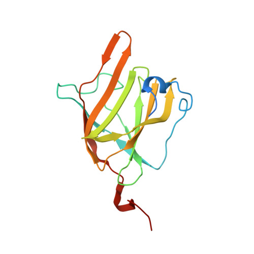
The human pathogen Staphylococcus aureus acquires heme iron from hemoglobin (Hb) via the action of a series of iron-regulated surface determinant (Isd) proteins. The cell wall anchored IsdB protein is recognized as the predominant Hb receptor, and is comprised of two NEAr transporter (NEAT) domains that act in concert to bind, extract, and transfer heme from Hb to downstream Isd proteins. Structural details of the NEAT 2 domain of IsdB have been investigated, but the molecular coordination between NEAT 2 and NEAT 1 to extract heme from hemoglobin has yet to be characterized. To obtain a more complete understanding of IsdB structure and function, we have solved the 3D solution structure of the NEAT 1 domain of IsdB (IsdB(N1)) spanning residues 125-272 of the full-length protein by NMR. The structure reveals a canonical NEAT domain fold and has particular structural similarity to the NEAT 1 and NEAT 2 domains of IsdH, which also interact with Hb. IsdB(N1) is also comprised of a short N-terminal helix, which has not been previously observed in other NEAT domain structures. Interestingly, the Hb binding region (loop 2 of IsdB(N1)) is disordered in solution. Analysis of Hb binding demonstrates that IsdB(N1) can bind metHb weakly and the affinity of this interaction is further increased by the presence of IsdB linker domain. IsdB(N1) loop 2 variants reveal that phenylalanine 164 (F164) of IsdB is necessary for Hb binding and rapid heme transfer from metHb to IsdB. Together, these findings provide a structural role for IsdB(N1) in enhancing the rate of extraction of metHb heme by the IsdB NEAT 2 domain.
Organizational Affiliation:
Department of Chemistry and Biochemistry, Montana State University , 103 Chemistry and Biochemistry Building, PO Box 173400, Bozeman, Montana 59717-3400, United States.