2AVU

Structure of the Escherichia coli FlhDC complex, a prokaryotic heteromeric regulator of transcription


Experimental Data Snapshot

  • Method: X-RAY DIFFRACTION
  • Resolution: 3.00 Å
  • R-Value Free: 0.255 
  • R-Value Work: 0.217 
  • R-Value Observed: 0.217 

wwPDB Validation   3D Report Full Report


This is version 1.3 of the entry. See complete history


Literature

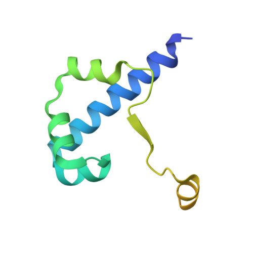
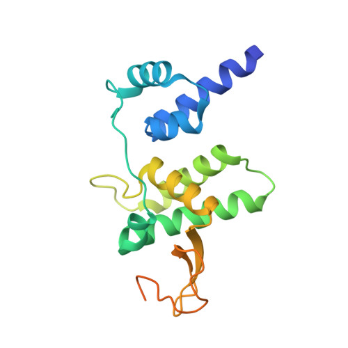
Structure of the Escherichia coli FlhDC Complex, a Prokaryotic Heteromeric Regulator of Transcription.

Wang, S.Fleming, R.T.Westbrook, E.M.Matsumura, P.McKay, D.B.

(2006) J Mol Biol 355: 798-808

  • DOI: https://doi.org/10.1016/j.jmb.2005.11.020
  • Primary Citation of Related Structures:  
    2AVU

  • PubMed Abstract: 

    The hetero-oligomeric complex of the FlhD and FlhC proteins (FlhDC) regulates transcription from several flagellar and non-flagellar operons in bacteria. The crystallographic structure of the Escherichia coli FlhDC complex has been solved to 3.0 A resolution, revealing a hexameric FlhD4FlhC2 assembly. In the complex, each FlhC protomer binds an FlhD2 dimer; the conformation of the dimer in the complex differs significantly from its conformation in the absence of FlhC. FlhC has a novel tertiary fold that includes a heretofore unrecognized zinc-binding site in which the ion is ligated by four cysteine residues. Gel shift experiments show that binding of the FlhDC complex to a cognate promoter bends the DNA by approximately 111 degrees . The structure of the FlhDC complex is compatible with models in which a fragment of operator DNA, at least 48 base-pairs in length, wraps around the complex and bends significantly when binding.


  • Organizational Affiliation

    Department of Structural Biology, Stanford University School of Medicine, Stanford, CA 94305, USA.


Macromolecules
Find similar proteins by:  (by identity cutoff)  |  3D Structure
Entity ID: 1
MoleculeChains Sequence LengthOrganismDetailsImage
Transcriptional activator flhD
A, B, C, D
116Escherichia coliMutation(s): 0 
Gene Names: flhDflbB
UniProt
Find proteins for P0A8S9 (Escherichia coli (strain K12))
Explore P0A8S9 
Go to UniProtKB:  P0A8S9
Entity Groups  
Sequence Clusters30% Identity50% Identity70% Identity90% Identity95% Identity100% Identity
UniProt GroupP0A8S9
Sequence Annotations
Expand
  • Reference Sequence
Find similar proteins by:  (by identity cutoff)  |  3D Structure
Entity ID: 2
MoleculeChains Sequence LengthOrganismDetailsImage
Flagellar transcriptional activator flhC
E, F
192Escherichia coliMutation(s): 0 
Gene Names: flhCflaI
UniProt
Find proteins for P0ABY7 (Escherichia coli (strain K12))
Explore P0ABY7 
Go to UniProtKB:  P0ABY7
Entity Groups  
Sequence Clusters30% Identity50% Identity70% Identity90% Identity95% Identity100% Identity
UniProt GroupP0ABY7
Sequence Annotations
Expand
  • Reference Sequence
Experimental Data & Validation

Experimental Data

  • Method: X-RAY DIFFRACTION
  • Resolution: 3.00 Å
  • R-Value Free: 0.255 
  • R-Value Work: 0.217 
  • R-Value Observed: 0.217 
  • Space Group: P 61
Unit Cell:
Length ( Å )Angle ( ˚ )
a = 151.137α = 90
b = 151.137β = 90
c = 114.16γ = 120
Software Package:
Software NamePurpose
CNSrefinement
HKL-2000data reduction
SCALEPACKdata scaling
SOLVEphasing

Structure Validation

View Full Validation Report


Entry History 

Deposition Data

Revision History  (Full details and data files)

  • Version 1.0: 2005-12-13
    Type: Initial release
  • Version 1.1: 2008-05-01
    Changes: Version format compliance
  • Version 1.2: 2011-07-13
    Changes: Version format compliance
  • Version 1.3: 2024-02-14
    Changes: Data collection, Database references, Derived calculations