1YBT

MYCOBACTERIUM TUBERCULOSIS ADENYLYL CYCLASE, RV1900C CHD


Experimental Data Snapshot

  • Method: X-RAY DIFFRACTION
  • Resolution: 2.31 Å
  • R-Value Free: 0.230 
  • R-Value Work: 0.194 
  • R-Value Observed: 0.194 

wwPDB Validation   3D Report Full Report


This is version 1.2 of the entry. See complete history


Literature

Origin of asymmetry in adenylyl cyclases: structures of Mycobacterium tuberculosis Rv1900c.

Sinha, S.C.Wetterer, M.Sprang, S.R.Schultz, J.E.Linder, J.U.

(2005) EMBO J 24: 663-673

  • DOI: https://doi.org/10.1038/sj.emboj.7600573
  • Primary Citation of Related Structures:  
    1YBT, 1YBU

  • PubMed Abstract: 

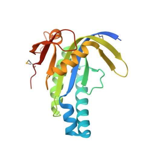
    Rv1900c, a Mycobacterium tuberculosis adenylyl cyclase, is composed of an N-terminal alpha/beta-hydrolase domain and a C-terminal cyclase homology domain. It has an unusual 7% guanylyl cyclase side-activity. A canonical substrate-defining lysine and a catalytic asparagine indispensable for mammalian adenylyl cyclase activity correspond to N342 and H402 in Rv1900c. Mutagenic analysis indicates that these residues are dispensable for activity of Rv1900c. Structures of the cyclase homology domain, solved to 2.4 A both with and without an ATP analog, form isologous, but asymmetric homodimers. The noncanonical N342 and H402 do not interact with the substrate. Subunits of the unliganded open dimer move substantially upon binding substrate, forming a closed dimer similar to the mammalian cyclase heterodimers, in which one interfacial active site is occupied and the quasi-dyad-related active site is occluded. This asymmetry indicates that both active sites cannot simultaneously be catalytically active. Such a mechanism of half-of-sites-reactivity suggests that mammalian heterodimeric adenylyl cyclases may have evolved from gene duplication of a primitive prokaryote-type cyclase, followed by loss of function in one active site.


  • Organizational Affiliation

    Howard Hughes Medical Institute and Department of Biochemistry, The University of Texas Southwestern Medical Center, Dallas, TX, USA.


Macromolecules
Find similar proteins by:  (by identity cutoff)  |  3D Structure
Entity ID: 1
MoleculeChains Sequence LengthOrganismDetailsImage
hydrolase, alpha/beta hydrolase fold family
A, B, C, D
184Mycobacterium tuberculosis CDC1551Mutation(s): 4 
Gene Names: Rv1900c
EC: 4.6.1.1
UniProt
Find proteins for O07732 (Mycobacterium tuberculosis (strain ATCC 25618 / H37Rv))
Explore O07732 
Go to UniProtKB:  O07732
Entity Groups  
Sequence Clusters30% Identity50% Identity70% Identity90% Identity95% Identity100% Identity
UniProt GroupO07732
Sequence Annotations
Expand
  • Reference Sequence
Small Molecules
Modified Residues  1 Unique
IDChains TypeFormula2D DiagramParent
MSE
Query on MSE
A, B, C, D
L-PEPTIDE LINKINGC5 H11 N O2 SeMET
Experimental Data & Validation

Experimental Data

  • Method: X-RAY DIFFRACTION
  • Resolution: 2.31 Å
  • R-Value Free: 0.230 
  • R-Value Work: 0.194 
  • R-Value Observed: 0.194 
  • Space Group: P 1 2 1
Unit Cell:
Length ( Å )Angle ( ˚ )
a = 90.704α = 90
b = 44.386β = 90
c = 80.425γ = 90
Software Package:
Software NamePurpose
CNSrefinement
HKL-2000data reduction
SCALEPACKdata scaling
SOLVEphasing

Structure Validation

View Full Validation Report


Entry History 

Deposition Data

Revision History  (Full details and data files)

  • Version 1.0: 2005-02-15
    Type: Initial release
  • Version 1.1: 2008-04-30
    Changes: Version format compliance
  • Version 1.2: 2011-07-13
    Changes: Source and taxonomy, Version format compliance