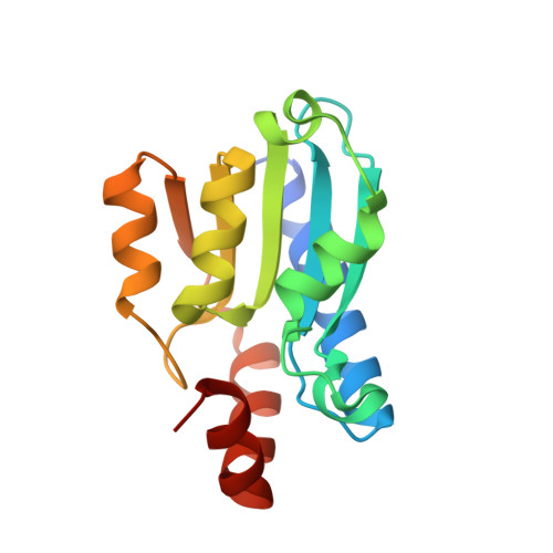
Crystal structure of yeast cytosine deaminase. Insights into enzyme mechanism and evolution
Ko, T.-P., Lin, J.-J., Hu, C.-Y., Hsu, Y.-H., Wang, A.H.-J., Liaw, S.-H.(2003) J Biol Chem 278: 19111-19117
- PubMed: 12637534
- DOI: https://doi.org/10.1074/jbc.M300874200
- Primary Citation of Related Structures:
1UAQ - PubMed Abstract:
Yeast cytosine deaminase is an attractive candidate for anticancer gene therapy because it catalyzes the deamination of the prodrug 5-fluorocytosine to form 5-fluorouracil. We report here the crystal structure of the enzyme in complex with the inhibitor 2-hydroxypyrimidine at 1.6-A resolution. The protein forms a tightly packed dimer with an extensive interface of 1450 A2 per monomer. The inhibitor was converted into a hydrated adduct as a transition-state analog. The essential zinc ion is ligated by the 4-hydroxyl group of the inhibitor together with His62, Cys91, and Cys94 from the protein. The enzyme shares similar active-site architecture to cytidine deaminases and an unusually high structural homology to 5-aminoimidazole-4-carboxamide-ribonucleotide transformylase and thereby may define a new superfamily. The unique C-terminal tail is involved in substrate specificity and also functions as a gate controlling access to the active site. The complex structure reveals a closed conformation, suggesting that substrate binding seals the active-site entrance so that the catalytic groups are sequestered from solvent. A comparison of the crystal structures of the bacterial and fungal cytosine deaminases provides an elegant example of convergent evolution, where starting from unrelated ancestral proteins, the same metal-assisted deamination is achieved through opposite chiral intermediates within distinctly different active sites.
Organizational Affiliation:
Institute of Biological Chemistry, Academia Sinica, Taipei 11529, Taiwan.