1TR8

Crystal Structure of archaeal Nascent Polypeptide-associated Complex (aeNAC)


Experimental Data Snapshot

  • Method: X-RAY DIFFRACTION
  • Resolution: 2.27 Å
  • R-Value Free: 0.240 
  • R-Value Work: 0.194 
  • R-Value Observed: 0.196 

wwPDB Validation   3D Report Full Report


This is version 1.2 of the entry. See complete history


Literature

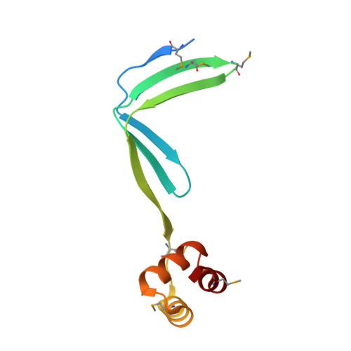
The crystal structure of archaeal nascent polypeptide-associated complex (NAC) reveals a unique fold and the presence of a UBA domain

Spreter, T.Pech, M.Beatrix, B.

(2005) J Biol Chem 280: 15849-15854

  • DOI: https://doi.org/10.1074/jbc.M500160200
  • Primary Citation of Related Structures:  
    1TR8

  • PubMed Abstract: 

    Nascent polypeptide-associated complex (NAC) was identified in eukaryotes as the first cytosolic factor that contacts the nascent polypeptide chain emerging from the ribosome. NAC is highly conserved from yeast to humans. Mutations in NAC cause severe embryonically lethal phenotypes in mice, Drosophila, and Caenorhabditis elegans. NAC was suggested to protect the nascent chain from inappropriate early interactions with cytosolic factors. Eukaryotic NAC is a heterodimer with two subunits sharing substantial homology with each other. All sequenced archaebacterial genomes exhibit only one gene homologous to the NAC subunits. Here we present the first archaebacterial NAC homolog. It forms a homodimer, and as eukaryotic NAC it is associated with ribosomes and contacts the emerging nascent chain on the ribosome. We present the first crystal structure of a NAC protein revealing two structural features: (i) a novel unique protein fold that mediates dimerization of the complex, and (ii) a ubiquitin-associated domain that suggests a yet unidentified role for NAC in the cellular protein quality control system via the ubiquitination pathway. Based on the presented structure we propose a model for the eukaryotic heterodimeric NAC domain.


  • Organizational Affiliation

    Institute for Chemistry-Crystallography, Free University of Berlin, Takustrasse 6, D-14195 Berlin, Germany.


Macromolecules
Find similar proteins by:  (by identity cutoff)  |  3D Structure
Entity ID: 1
MoleculeChains Sequence LengthOrganismDetailsImage
conserved protein (MTH177)
A, B
102Methanothermobacter marburgensisMutation(s): 9 
Gene Names: MTH177 (amonoacids 19-117)
UniProt
Find proteins for P0C0K9 (Methanothermobacter marburgensis (strain ATCC BAA-927 / DSM 2133 / JCM 14651 / NBRC 100331 / OCM 82 / Marburg))
Explore P0C0K9 
Go to UniProtKB:  P0C0K9
Entity Groups  
Sequence Clusters30% Identity50% Identity70% Identity90% Identity95% Identity100% Identity
UniProt GroupP0C0K9
Sequence Annotations
Expand
  • Reference Sequence
Small Molecules
Modified Residues  1 Unique
IDChains TypeFormula2D DiagramParent
MSE
Query on MSE
A, B
L-PEPTIDE LINKINGC5 H11 N O2 SeMET
Experimental Data & Validation

Experimental Data

  • Method: X-RAY DIFFRACTION
  • Resolution: 2.27 Å
  • R-Value Free: 0.240 
  • R-Value Work: 0.194 
  • R-Value Observed: 0.196 
  • Space Group: P 43 21 2
Unit Cell:
Length ( Å )Angle ( ˚ )
a = 90.89α = 90
b = 90.89β = 90
c = 49.476γ = 90
Software Package:
Software NamePurpose
REFMACrefinement
MOSFLMdata reduction
CCP4data scaling
PHENIXphasing
SHARPphasing
RESOLVEphasing

Structure Validation

View Full Validation Report


Entry History 

Deposition Data

Revision History  (Full details and data files)

  • Version 1.0: 2005-02-01
    Type: Initial release
  • Version 1.1: 2008-04-30
    Changes: Version format compliance
  • Version 1.2: 2011-07-13
    Changes: Advisory, Version format compliance