1SHX

Ephrin A5 ligand structure


Experimental Data Snapshot

  • Method: X-RAY DIFFRACTION
  • Resolution: 2.10 Å
  • R-Value Free: 0.278 
  • R-Value Work: 0.224 

wwPDB Validation   3D Report Full Report


This is version 2.1 of the entry. See complete history


Literature

Three distinct molecular surfaces in ephrin-A5 are essential for a functional interaction with EphA3.

Day, B.To, C.Himanen, J.P.Smith, F.M.Nikolov, D.B.Boyd, A.W.Lackmann, M.

(2005) J Biol Chem 280: 26526-26532

  • DOI: https://doi.org/10.1074/jbc.M504972200
  • Primary Citation of Related Structures:  
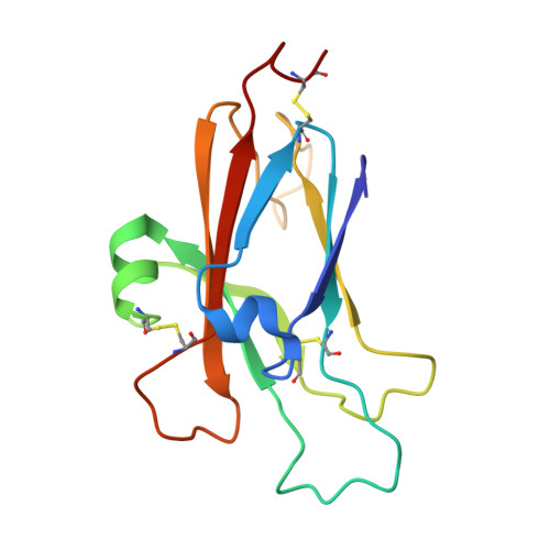
    1SHX

  • PubMed Abstract: 

    Eph receptor tyrosine kinases (Ephs) function as molecular relays that interact with cell surface-bound ephrin ligands to direct the position of migrating cells. Structural studies revealed that, through two distinct contact surfaces on opposite sites of each protein, Eph and ephrin binding domains assemble into symmetric, circular heterotetramers. However, Eph signal initiation requires the assembly of higher order oligomers, suggesting additional points of contact. By screening a random library of EphA3 binding-compromised ephrin-A5 mutants, we have now determined ephrin-A5 residues that are essential for the assembly of high affinity EphA3 signaling complexes. In addition to the two interfaces predicted from the crystal structure of the homologous EphB2.ephrin-B2 complex, we identified a cluster of 10 residues on the ephrin-A5 E alpha-helix, the E-F loop, the underlying H beta-strand, as well as the nearby B-C loop, which define a distinct third surface required for oligomerization and activation of EphA3 signaling. Together with a corresponding third surface region identified recently outside of the minimal ephrin binding domain of EphA3, our findings provide experimental evidence for the essential contribution of three distinct protein-interaction interfaces to assemble functional EphA3 signaling complexes.


  • Organizational Affiliation

    Queensland Institute of Medical Research, P. O. Royal Brisbane Hospital 4029, Queensland, Australia.


Macromolecules
Find similar proteins by:  (by identity cutoff)  |  3D Structure
Entity ID: 1
MoleculeChains Sequence LengthOrganismDetailsImage
Ephrin-A5
A, B
138Mus musculusMutation(s): 0 
Gene Names: EFNA5EPLG7LERK7EPL7
UniProt & NIH Common Fund Data Resources
Find proteins for O08543 (Mus musculus)
Explore O08543 
Go to UniProtKB:  O08543
IMPC:  MGI:107444
Entity Groups  
Sequence Clusters30% Identity50% Identity70% Identity90% Identity95% Identity100% Identity
UniProt GroupO08543
Sequence Annotations
Expand
  • Reference Sequence
Oligosaccharides

Help

Entity ID: 2
MoleculeChains Length2D Diagram Glycosylation3D Interactions
2-acetamido-2-deoxy-beta-D-glucopyranose-(1-4)-2-acetamido-2-deoxy-beta-D-glucopyranose-(1-4)-2-acetamido-2-deoxy-beta-D-glucopyranose
C
3N-Glycosylation
Glycosylation Resources
GlyTouCan:  G47362BJ
GlyCosmos:  G47362BJ
GlyGen:  G47362BJ
Entity ID: 3
MoleculeChains Length2D Diagram Glycosylation3D Interactions
2-acetamido-2-deoxy-beta-D-glucopyranose-(1-4)-2-acetamido-2-deoxy-beta-D-glucopyranose
D
2N-Glycosylation
Glycosylation Resources
GlyTouCan:  G42666HT
GlyCosmos:  G42666HT
GlyGen:  G42666HT
Biologically Interesting Molecules (External Reference) 1 Unique
Experimental Data & Validation

Experimental Data

  • Method: X-RAY DIFFRACTION
  • Resolution: 2.10 Å
  • R-Value Free: 0.278 
  • R-Value Work: 0.224 
  • Space Group: P 1 21 1
Unit Cell:
Length ( Å )Angle ( ˚ )
a = 36.21α = 90
b = 70.08β = 95.94
c = 58.46γ = 90
Software Package:
Software NamePurpose
DENZOdata reduction
SCALEPACKdata scaling
AMoREphasing
CNSrefinement

Structure Validation

View Full Validation Report


Entry History 

Deposition Data

Revision History  (Full details and data files)

  • Version 1.0: 2005-04-19
    Type: Initial release
  • Version 1.1: 2008-04-29
    Changes: Version format compliance
  • Version 1.2: 2011-07-13
    Changes: Non-polymer description, Version format compliance
  • Version 2.0: 2020-07-29
    Type: Remediation
    Reason: Carbohydrate remediation
    Changes: Atomic model, Data collection, Derived calculations, Structure summary
  • Version 2.1: 2024-04-03
    Changes: Data collection, Database references, Refinement description, Structure summary