1VSG

2.9 ANGSTROMS RESOLUTION STRUCTURE OF THE N-TERMINAL DOMAIN OF A VARIANT SURFACE GLYCOPROTEIN FROM TRYPANOSOMA BRUCEI


Experimental Data Snapshot

  • Method: X-RAY DIFFRACTION
  • Resolution: 2.90 Å
  • R-Value Work: 0.220 

wwPDB Validation   3D Report Full Report


This is version 2.0 of the entry. See complete history

Re-refinement Note

A newer entry is available that reflects an alternative modeling of the original data: 7P56


Literature

2.9 A resolution structure of the N-terminal domain of a variant surface glycoprotein from Trypanosoma brucei.

Freymann, D.Down, J.Carrington, M.Roditi, I.Turner, M.Wiley, D.

(1990) J Mol Biol 216: 141-160

  • DOI: https://doi.org/10.1016/S0022-2836(05)80066-X
  • Primary Citation of Related Structures:  
    1VSG

  • PubMed Abstract: 

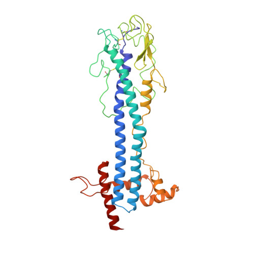
    The variant surface glycoprotein (VSG) of Trypanosoma brucei forms a coat on the surface of the parasite; by the expression of a series of antigenically distinct VSGs in the surface coat the parasite escapes the host immune response. The 2.9 A resolution crystal structure of the N-terminal domain of one variant, MITat 1.2, has been determined. The structure was solved using data collected from two crystal forms. Initially a partial model was built into an electron density map based on multiple isomorphous replacement phases and improved by phase combination methods. Subsequently this model was used to obtain the molecular replacement solution for a second crystal form, providing starting phases which were refined using 2-fold non-crystallographic symmetry averaging. The current model includes 362 residues and has been refined using X-PLOR to an R value of 0.22 for data between 7 and 2.9 A. The molecule is a dimer, approximately 100 A long, having an asymmetrical cross section with maximum dimensions of approximately 40 A x 60 A. Two long, approximately 70 A, alpha-helices from each monomer pack together to form, with several other helices, a core helix bundle that extends nearly the full length of the molecule. The "top" of the protein, which in the surface coat may be exposed to the external environment, is formed from the ends of the two long helices, a short three-stranded beta-sheet, and a strand having irregular conformation that packs above these secondary structure elements. Two conserved disulfide bridges are in this part of the molecule. Several elements of the MITat 1.2 sequence, which contribute to the formation of the helix bundle structure, have been identified. These elements can be found in the sequences of several different VSGs, suggesting that to some extent the VSG structure is conserved in those variants.


  • Organizational Affiliation

    Department of Biochemistry and Molecular Biology, Harvard University, Cambridge, MA 02138.


Macromolecules
Find similar proteins by:  (by identity cutoff)  |  3D Structure
Entity ID: 1
MoleculeChains Sequence LengthOrganismDetailsImage
VARIANT SURFACE GLYCOPROTEIN MITAT 1.2
A, B
364Trypanosoma brucei bruceiMutation(s): 0 
Membrane Entity: Yes 
UniProt
Find proteins for P26332 (Trypanosoma brucei brucei)
Explore P26332 
Go to UniProtKB:  P26332
Entity Groups  
Sequence Clusters30% Identity50% Identity70% Identity90% Identity95% Identity100% Identity
UniProt GroupP26332
Sequence Annotations
Expand
  • Reference Sequence
Oligosaccharides

Help

Entity ID: 2
MoleculeChains Length2D Diagram Glycosylation3D Interactions
beta-D-mannopyranose-(1-4)-2-acetamido-2-deoxy-beta-D-glucopyranose-(1-4)-2-acetamido-2-deoxy-beta-D-glucopyranose
C
3N-Glycosylation
Glycosylation Resources
GlyTouCan:  G15407YE
GlyCosmos:  G15407YE
GlyGen:  G15407YE
Entity ID: 3
MoleculeChains Length2D Diagram Glycosylation3D Interactions
alpha-D-mannopyranose-(1-4)-2-acetamido-2-deoxy-beta-D-glucopyranose-(1-4)-2-acetamido-2-deoxy-beta-D-glucopyranose
D
3N-Glycosylation
Glycosylation Resources
GlyTouCan:  G62182OO
GlyCosmos:  G62182OO
GlyGen:  G62182OO
Experimental Data & Validation

Experimental Data

  • Method: X-RAY DIFFRACTION
  • Resolution: 2.90 Å
  • R-Value Work: 0.220 
  • Space Group: P 21 21 21
Unit Cell:
Length ( Å )Angle ( ˚ )
a = 95.5α = 90
b = 97.5β = 90
c = 123.7γ = 90
Software Package:
Software NamePurpose
X-PLORrefinement

Structure Validation

View Full Validation Report


Entry History 

Deposition Data

Revision History  (Full details and data files)

  • Version 1.0: 1992-01-15
    Type: Initial release
  • Version 1.1: 2008-03-24
    Changes: Version format compliance
  • Version 1.2: 2011-07-13
    Changes: Non-polymer description, Version format compliance
  • Version 2.0: 2020-07-29
    Type: Remediation
    Reason: Carbohydrate remediation
    Changes: Advisory, Atomic model, Data collection, Derived calculations, Other, Structure summary