An examination of coaxial stacking of helical stems in a pseudoknot motif: the gene 32 messenger RNA pseudoknot of bacteriophage T2.
Holland, J.A., Hansen, M.R., Du, Z., Hoffman, D.W.(1999) RNA 5: 257-271
- PubMed: 10024177
- DOI: https://doi.org/10.1017/s1355838299981360
- Primary Citation of Related Structures:
2TPK - PubMed Abstract:
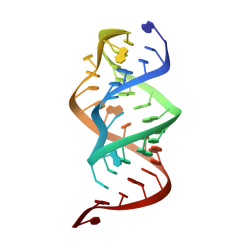
The RNA pseudoknot located at the 5' end of the gene 32 messenger RNA of bacteriophage T2 contains two A-form helical stems connected by two loops, in an H-type pseudoknot topology. A combination of multidimensional NMR methods and isotope labeling were used to investigate the pseudoknot structure, resulting in a more detailed structural model than provided by earlier homonuclear NMR studies. Of particular significance, the interface between the stacked helical stems within the pseudoknot motif is described in detail. The two stems are stacked in a coaxial manner, with an approximately 18 degrees rotation of stem1 relative to stem2 about an axis that is parallel to the helical axis. This rotation serves to relieve what would otherwise be a relatively close phosphate-phosphate contact at the junction of the two stems, while preserving the stabilizing effects of base stacking. The ability of the NMR data to determine pseudoknot bending was critically assessed. The data were found to be a modestly precise indicator of pseudoknot bending, with the angle between the helical axes of stem1 and stem2 being in the range of 15+/-15 degrees. Pseudoknot models with bend angles within this range are equally consistent with the data, since they differ by only small amounts in the relatively short-range interproton distances from which the structure was derived. The gene 32 messenger RNA pseudoknot was compared with other RNA structures with coaxial or near-coaxial stacked helical stems.
Organizational Affiliation:
Department of Chemistry and Biochemistry, Institute for Cell and Molecular Biology, University of Texas at Austin, 78712, USA.